7WLJ

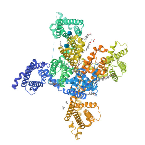
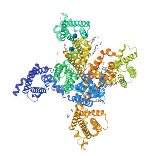
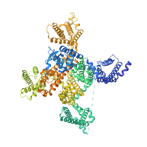
CryoEM structure of human low-voltage activated T-type calcium channel Cav3.3 in complex with mibefradil (MIB)


Experimental Data Snapshot

  • Method: ELECTRON MICROSCOPY
  • Resolution: 3.90 Å
  • Aggregation State: PARTICLE 
  • Reconstruction Method: SINGLE PARTICLE 

wwPDB Validation   3D Report Full Report


This is version 1.2 of the entry. See complete history


Literature

Structure, gating, and pharmacology of human Ca V 3.3 channel.

He, L.Yu, Z.Geng, Z.Huang, Z.Zhang, C.Dong, Y.Gao, Y.Wang, Y.Chen, Q.Sun, L.Ma, X.Huang, B.Wang, X.Zhao, Y.

(2022) Nat Commun 13: 2084-2084

  • DOI: https://doi.org/10.1038/s41467-022-29728-0
  • Primary Citation of Related Structures:  
    7WLI, 7WLJ, 7WLK, 7WLL

  • PubMed Abstract: 

    The low-voltage activated T-type calcium channels regulate cellular excitability and oscillatory behavior of resting membrane potential which trigger many physiological events and have been implicated with many diseases. Here, we determine structures of the human T-type Ca V 3.3 channel, in the absence and presence of antihypertensive drug mibefradil, antispasmodic drug otilonium bromide and antipsychotic drug pimozide. Ca V 3.3 contains a long bended S6 helix from domain III, with a positive charged region protruding into the cytosol, which is critical for T-type Ca V channel activation at low voltage. The drug-bound structures clearly illustrate how these structurally different compounds bind to the same central cavity inside the Ca V 3.3 channel, but are mediated by significantly distinct interactions between drugs and their surrounding residues. Phospholipid molecules penetrate into the central cavity in various extent to shape the binding pocket and play important roles in stabilizing the inhibitor. These structures elucidate mechanisms of channel gating, drug recognition, and actions, thus pointing the way to developing potent and subtype-specific drug for therapeutic treatments of related disorders.


  • Organizational Affiliation

    National Laboratory of Biomacromolecules, CAS Center for Excellence in Biomacromolecules, Institute of Biophysics, Chinese Academy of Sciences, Beijing, 100101, China.


Macromolecules
Find similar proteins by:  (by identity cutoff)  |  3D Structure
Entity ID: 1
MoleculeChains Sequence LengthOrganismDetailsImage
Voltage-dependent T-type calcium channel subunit alpha-1I2,223Homo sapiensMutation(s): 0 
Gene Names: CACNA1IKIAA1120
Membrane Entity: Yes 
UniProt & NIH Common Fund Data Resources
Find proteins for Q9P0X4 (Homo sapiens)
Explore Q9P0X4 
Go to UniProtKB:  Q9P0X4
PHAROS:  Q9P0X4
GTEx:  ENSG00000100346 
Entity Groups  
Sequence Clusters30% Identity50% Identity70% Identity90% Identity95% Identity100% Identity
UniProt GroupQ9P0X4
Glycosylation
Glycosylation Sites: 2Go to GlyGen: Q9P0X4-1
Sequence Annotations
Expand
  • Reference Sequence
Small Molecules
Ligands 5 Unique
IDChains Name / Formula / InChI Key2D Diagram3D Interactions
3PE
Query on 3PE

Download Ideal Coordinates CCD File 
C [auth A],
D [auth A],
F [auth A],
G [auth A],
H [auth A]
1,2-Distearoyl-sn-glycerophosphoethanolamine
C41 H82 N O8 P
LVNGJLRDBYCPGB-LDLOPFEMSA-N
MWV (Subject of Investigation/LOI)
Query on MWV

Download Ideal Coordinates CCD File 
B [auth A](1S,2S)-2-(2-{[3-(1H-benzimidazol-2-yl)propyl](methyl)amino}ethyl)-6-fluoro-1-(propan-2-yl)-1,2,3,4-tetrahydronaphthalen-2-yl methoxyacetate
C29 H38 F N3 O3
HBNPJJILLOYFJU-VMPREFPWSA-N
Y01
Query on Y01

Download Ideal Coordinates CCD File 
I [auth A]
J [auth A]
L [auth A]
N [auth A]
O [auth A]
I [auth A],
J [auth A],
L [auth A],
N [auth A],
O [auth A],
P [auth A]
CHOLESTEROL HEMISUCCINATE
C31 H50 O4
WLNARFZDISHUGS-MIXBDBMTSA-N
NAG
Query on NAG

Download Ideal Coordinates CCD File 
K [auth A],
M [auth A]
2-acetamido-2-deoxy-beta-D-glucopyranose
C8 H15 N O6
OVRNDRQMDRJTHS-FMDGEEDCSA-N
CA
Query on CA

Download Ideal Coordinates CCD File 
E [auth A]CALCIUM ION
Ca
BHPQYMZQTOCNFJ-UHFFFAOYSA-N
Binding Affinity Annotations 
IDSourceBinding Affinity
MWV BindingDB:  7WLJ IC50: min: 126, max: 1000 (nM) from 3 assay(s)
Experimental Data & Validation

Experimental Data

  • Method: ELECTRON MICROSCOPY
  • Resolution: 3.90 Å
  • Aggregation State: PARTICLE 
  • Reconstruction Method: SINGLE PARTICLE 

Structure Validation

View Full Validation Report


Entry History & Funding Information

Deposition Data


Funding OrganizationLocationGrant Number
Chinese Academy of SciencesChina--

Revision History  (Full details and data files)

  • Version 1.0: 2022-05-04
    Type: Initial release
  • Version 1.1: 2022-05-25
    Changes: Database references
  • Version 1.2: 2024-11-06
    Changes: Data collection, Structure summary