7WJ4 | pdb_00007wj4

Structural basis for ligand binding modes of CTP synthase


Experimental Data Snapshot

  • Method: ELECTRON MICROSCOPY
  • Resolution: 3.15 Å
  • Aggregation State: FILAMENT 
  • Reconstruction Method: SINGLE PARTICLE 

wwPDB Validation 3D Report Full Report

Validation slider image for 7WJ4

This is version 1.1 of the entry. See complete history

Literature

Structural basis for ligand binding modes of CTP synthase.

Zhou, X.Guo, C.J.Chang, C.C.Zhong, J.Hu, H.H.Lu, G.M.Liu, J.L.

(2021) Proc Natl Acad Sci U S A 118

  • DOI: https://doi.org/10.1073/pnas.2026621118
  • Primary Citation Related Structures: 
    7DPT, 7DPW, 7WIZ, 7WJ4

  • PubMed Abstract: 

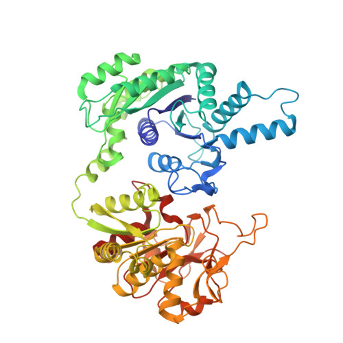
    Cytidine triphosphate synthase (CTPS), which comprises an ammonia ligase domain and a glutamine amidotransferase domain, catalyzes the final step of de novo CTP biosynthesis. The activity of CTPS is regulated by the binding of four nucleotides and glutamine. While glutamine serves as an ammonia donor for the ATP-dependent conversion of UTP to CTP, the fourth nucleotide GTP acts as an allosteric activator. Models have been proposed to explain the mechanisms of action at the active site of the ammonia ligase domain and the conformational changes derived by GTP binding. However, actual GTP/ATP/UTP binding modes and relevant conformational changes have not been revealed fully. Here, we report the discovery of binding modes of four nucleotides and a glutamine analog 6-diazo-5-oxo-L-norleucine in Drosophila CTPS by cryo-electron microscopy with near-atomic resolution. Interactions between GTP and surrounding residues indicate that GTP acts to coordinate reactions at both domains by directly blocking ammonia leakage and stabilizing the ammonia tunnel. Additionally, we observe the ATP-dependent UTP phosphorylation intermediate and determine interacting residues at the ammonia ligase. A noncanonical CTP binding at the ATP binding site suggests another layer of feedback inhibition. Our findings not only delineate the structure of CTPS in the presence of all substrates but also complete our understanding of the underlying mechanisms of the allosteric regulation and CTP synthesis.


  • Organizational Affiliation
    • School of Life Science and Technology, ShanghaiTech University, Shanghai, 201210, China.

Macromolecule Content 

  • Total Structure Weight: 256.71 kDa 
  • Atom Count: 17,960 
  • Modeled Residue Count: 2,224 
  • Deposited Residue Count: 2,224 
  • Unique protein chains: 1

Macromolecules

Find similar proteins by:|  3D Structure
Entity ID: 1
MoleculeChains  Sequence LengthOrganismDetailsImage
CTP synthaseA [auth D],
B [auth A],
C [auth B],
D [auth C]
556Drosophila melanogasterMutation(s): 0 
Gene Names: CTPsynCG45070
EC: 6.3.4.2
UniProt
Find proteins for Q9VUL1 (Drosophila melanogaster)
Explore Q9VUL1 
Go to UniProtKB:  Q9VUL1
Entity Groups
Sequence Clusters30% Identity50% Identity70% Identity90% Identity95% Identity100% Identity
UniProt GroupQ9VUL1
Sequence Annotations
Expand
Reference Sequence

Small Molecules

Ligands 5 Unique
IDChains Name / Formula / InChI Key2D Diagram3D Interactions
GTP
(Subject of Investigation/LOI)

Query on GTP


Download:Ideal Coordinates CCD File
BA [auth C],
E [auth D],
N [auth A],
S [auth B]
GUANOSINE-5'-TRIPHOSPHATE
C10 H16 N5 O14 P3
XKMLYUALXHKNFT-UUOKFMHZSA-N
ATP
(Subject of Investigation/LOI)

Query on ATP


Download:Ideal Coordinates CCD File
CA [auth C],
F [auth D],
O [auth A],
T [auth B]
ADENOSINE-5'-TRIPHOSPHATE
C10 H16 N5 O13 P3
ZKHQWZAMYRWXGA-KQYNXXCUSA-N
UTP
(Subject of Investigation/LOI)

Query on UTP


Download:Ideal Coordinates CCD File
J [auth D],
L [auth A],
X [auth B],
Z [auth C]
URIDINE 5'-TRIPHOSPHATE
C9 H15 N2 O15 P3
PGAVKCOVUIYSFO-XVFCMESISA-N
GGL
(Subject of Investigation/LOI)

Query on GGL


Download:Ideal Coordinates CCD File
DA [auth C],
G [auth D],
P [auth A],
U [auth B]
GAMMA-L-GLUTAMIC ACID
C5 H9 N O4
WHUUTDBJXJRKMK-VKHMYHEASA-N
MG
(Subject of Investigation/LOI)

Query on MG


Download:Ideal Coordinates CCD File
AA [auth C]
EA [auth C]
FA [auth C]
H [auth D]
I [auth D]
AA [auth C],
EA [auth C],
FA [auth C],
H [auth D],
I [auth D],
K [auth D],
M [auth A],
Q [auth A],
R [auth A],
V [auth B],
W [auth B],
Y [auth B]
MAGNESIUM ION
Mg
JLVVSXFLKOJNIY-UHFFFAOYSA-N

Experimental Data & Validation

Experimental Data

  • Method: ELECTRON MICROSCOPY
  • Resolution: 3.15 Å
  • Aggregation State: FILAMENT 
  • Reconstruction Method: SINGLE PARTICLE 

Structure Validation

View Full Validation Report


Entry History 

& Funding Information

Deposition Data


Funding OrganizationLocationGrant Number
National Natural Science Foundation of China (NSFC)China--

Revision History  (Full details and data files)

  • Version 1.0: 2023-01-11
    Type: Initial release
  • Version 1.1: 2025-09-17
    Changes: Advisory, Data collection, Derived calculations, Structure summary