7WIK | pdb_00007wik

Crystal structure of oligoribonuclease of Mycobacterium smegmatis mc2 155


Experimental Data Snapshot

  • Method: X-RAY DIFFRACTION
  • Resolution: 1.87 Å
  • R-Value Free: 
    0.222 (Depositor), 0.227 (DCC) 
  • R-Value Work: 
    0.179 (Depositor), 0.188 (DCC) 
  • R-Value Observed: 
    0.181 (Depositor) 

Starting Model: experimental
View more details

wwPDB Validation 3D Report Full Report

Validation slider image for 7WIK

This is version 1.1 of the entry. See complete history

Literature

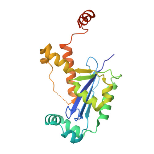
Three-dimensional structure of a mycobacterial oligoribonuclease reveals a unique C-terminal tail that stabilizes the homodimer.

Badhwar, P.Khan, S.H.Taneja, B.

(2022) J Biological Chem 298: 102595-102595

  • DOI: https://doi.org/10.1016/j.jbc.2022.102595
  • Primary Citation Related Structures: 
    7VH4, 7WIK

  • PubMed Abstract: 

    Oligoribonucleases (Orns) are highly conserved DnaQ-fold 3'-5' exoribonucleases that have been found to carry out the last step of cyclic-di-GMP (c-di-GMP) degradation, that is, pGpG to GMP in several bacteria. Removal of pGpG is critical for c-di-GMP homeostasis, as excess uncleaved pGpG can have feedback inhibition on phosphodiesterases, thereby perturbing cellular signaling pathways regulated by c-di-GMP. Perturbation of c-di-GMP levels not only affects survival under hypoxic, reductive stress, or nutrient-limiting conditions but also affects pathogenicity in infection models as well as antibiotic response in mycobacteria. Here, we have determined the crystal structure of MSMEG_4724, the Orn of Mycobacterium smegmatis (Ms_orn) to 1.87 Å resolution to investigate the function of its extended C-terminal tail that is unique among bacterial Orns. Ms_orn is a homodimer with the canonical RNase-H fold of exoribonucleases and conserved catalytic residues in the active site. Further examination of the substrate-binding site with a modeled pGpG emphasized the role of a phosphate cap and "3'OH cap" in constricting a 2-mer substrate in the active site. The unique C-terminal tail of Ms_orn aids dimerization by forming a handshake-like flap over the second protomer of the dimer. Our thermal and denaturant-induced unfolding experiments suggest that it helps in higher stability of Ms_orn as compared with Escherichia coli Orn or a C-terminal deletion mutant. We also show that the C-terminal tail is required for modulating response to stress agents in vivo. These results will help in further evaluating the role of signaling and regulation by c-di-GMP in mycobacteria.


  • Organizational Affiliation
    • CSIR-Institute of Genomics and Integrative Biology (CSIR-IGIB), New Delhi, India; Academy of Scientific and Innovative Research (AcSIR), Ghaziabad, India.

Macromolecule Content 

  • Total Structure Weight: 95.46 kDa 
  • Atom Count: 7,150 
  • Modeled Residue Count: 781 
  • Deposited Residue Count: 840 
  • Unique protein chains: 1

Macromolecules

Find similar proteins by:|  3D Structure
Entity ID: 1
MoleculeChains  Sequence LengthOrganismDetailsImage
Oligoribonuclease
A, B, C, D
210Mycolicibacterium smegmatis MC2 155Mutation(s): 0 
Gene Names: orn
EC: 3.1.13.3 (PDB Primary Data), 3.1 (UniProt)
UniProt
Find proteins for A0R1E6 (Mycolicibacterium smegmatis (strain ATCC 700084 / mc(2)155))
Explore A0R1E6 
Go to UniProtKB:  A0R1E6
Entity Groups
Sequence Clusters30% Identity50% Identity70% Identity90% Identity95% Identity100% Identity
UniProt GroupA0R1E6
Sequence Annotations
Expand
Reference Sequence

Small Molecules

Ligands 6 Unique
IDChains Name / Formula / InChI Key2D Diagram3D Interactions
PEG

Query on PEG


Download:Ideal Coordinates CCD File
CA [auth D]DI(HYDROXYETHYL)ETHER
C4 H10 O3
MTHSVFCYNBDYFN-UHFFFAOYSA-N
SO4

Query on SO4


Download:Ideal Coordinates CCD File
BA [auth C]
GA [auth D]
H [auth A]
HA [auth D]
IA [auth D]
BA [auth C],
GA [auth D],
H [auth A],
HA [auth D],
IA [auth D],
J [auth B],
N [auth B],
O [auth B],
Q [auth B],
R [auth C],
W [auth C],
X [auth C],
Y [auth C]
SULFATE ION
O4 S
QAOWNCQODCNURD-UHFFFAOYSA-L
GOL

Query on GOL


Download:Ideal Coordinates CCD File
I [auth A],
JA [auth D],
P [auth B],
Z [auth C]
GLYCEROL
C3 H8 O3
PEDCQBHIVMGVHV-UHFFFAOYSA-N
ACT

Query on ACT


Download:Ideal Coordinates CCD File
EA [auth D]
F [auth A]
FA [auth D]
G [auth A]
L [auth B]
EA [auth D],
F [auth A],
FA [auth D],
G [auth A],
L [auth B],
M [auth B],
T [auth C],
U [auth C],
V [auth C]
ACETATE ION
C2 H3 O2
QTBSBXVTEAMEQO-UHFFFAOYSA-M
K
(Subject of Investigation/LOI)

Query on K


Download:Ideal Coordinates CCD File
DA [auth D],
E [auth A],
K [auth B],
S [auth C]
POTASSIUM ION
K
NPYPAHLBTDXSSS-UHFFFAOYSA-N
MG

Query on MG


Download:Ideal Coordinates CCD File
AA [auth C]MAGNESIUM ION
Mg
JLVVSXFLKOJNIY-UHFFFAOYSA-N

Experimental Data & Validation

Experimental Data

  • Method: X-RAY DIFFRACTION
  • Resolution: 1.87 Å
  • R-Value Free:  0.222 (Depositor), 0.227 (DCC) 
  • R-Value Work:  0.179 (Depositor), 0.188 (DCC) 
  • R-Value Observed: 0.181 (Depositor) 
Space Group: P 21 21 21
Unit Cell:
Length ( Å )Angle ( ˚ )
a = 60.25α = 90
b = 97.26β = 90
c = 146.73γ = 90
Software Package:
Software NamePurpose
REFMACrefinement
XSCALEdata scaling
PDB_EXTRACTdata extraction
autoPROCdata reduction
PHASERphasing

Structure Validation

View Full Validation Report


Entry History 

& Funding Information

Deposition Data


Funding OrganizationLocationGrant Number
Not funded--

Revision History  (Full details and data files)

  • Version 1.0: 2022-12-21
    Type: Initial release
  • Version 1.1: 2023-11-29
    Changes: Data collection, Refinement description