7WG3 | pdb_00007wg3

Structural basis of interleukin-17B receptor in complex with a neutralizing antibody D9 for guiding humanization and affinity maturation for cancer therapy


Experimental Data Snapshot

  • Method: X-RAY DIFFRACTION
  • Resolution: 2.19 Å
  • R-Value Free: 
    0.238 (Depositor), 0.245 (DCC) 
  • R-Value Work: 
    0.193 (Depositor) 
  • R-Value Observed: 
    0.195 (Depositor) 

Starting Models: experimental
View more details

wwPDB Validation 3D Report Full Report

Validation slider image for 7WG3

Ligand Structure Quality Assessment 


This is version 1.2 of the entry. See complete history

Literature

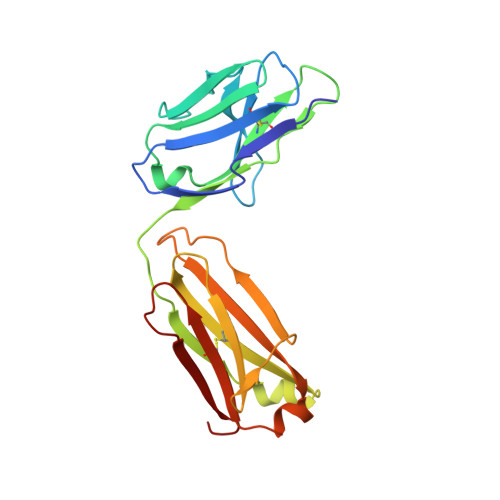
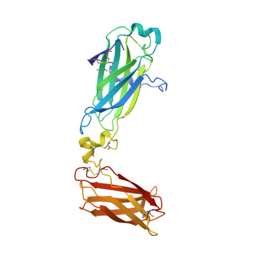
Structural basis of interleukin-17B receptor in complex with a neutralizing antibody for guiding humanization and affinity maturation.

Lee, W.H.Chen, X.Liu, I.J.Lee, J.H.Hu, C.M.Wu, H.C.Wang, S.K.Lee, W.H.Ma, C.

(2022) Cell Rep 41: 111555-111555

  • DOI: https://doi.org/10.1016/j.celrep.2022.111555
  • Primary Citation Related Structures: 
    7WG3

  • PubMed Abstract: 

    Upregulation of interleukin-17 receptor B (IL-17RB) is known to be oncogenic, while other IL-17 receptors and ligands are generally involved in pro-inflammatory pathways. We identify a mouse neutralizing monoclonal antibody (mAb) D9, which blocks the IL-17RB/IL-17B pathway and inhibits pancreatic tumorigenesis in an orthotopic mouse model. The X-ray crystal structure of the IL-17RB ectodomain in complex with its neutralizing antibody D9 shows that D9 binds to a predicted ligand binding interface and engages with the A'-A loop of IL-17RB fibronectin III domain 1 in a unique conformational state. This structure also provides important paratope information to guide the design of antibody humanization and affinity maturation of D9, resulting in a humanized 1B12 antibody with marginal affinity loss and effective neutralization of IL-17B/IL-17RB signaling to impede tumorigenesis in a mouse xenograft model.


  • Organizational Affiliation
    • Genomics Research Center, Academia Sinica, Taipei, Taiwan; Chemical Biology and Molecular Biophysics, Taiwan International Graduate Program, Academia Sinica, Taipei, Taiwan; Department of Chemistry, National Tsing Hua University, Hsinchu, Taiwan.

Macromolecule Content 

  • Total Structure Weight: 305.75 kDa 
  • Atom Count: 21,987 
  • Modeled Residue Count: 2,712 
  • Deposited Residue Count: 2,752 
  • Unique protein chains: 3

Macromolecules

Find similar proteins by:|  3D Structure
Entity ID: 1
MoleculeChains  Sequence LengthOrganismDetailsImage
Light chain of D9 Fab
A, B, C, D
213Mus musculusMutation(s): 0 
Find similar proteins by:|  3D Structure
Entity ID: 2
MoleculeChains  Sequence LengthOrganismDetailsImage
Heavy chain of D9 Fab
E, F, G, H
220Mus musculusMutation(s): 0 
Find similar proteins by:|  3D Structure
Entity ID: 3
MoleculeChains  Sequence LengthOrganismDetailsImage
IL17RB protein
I, J, K, L
255Bos taurusMutation(s): 0 
Gene Names: IL17RB
UniProt
Find proteins for A3KN55 (Bos taurus)
Explore A3KN55 
Go to UniProtKB:  A3KN55
Entity Groups
Sequence Clusters30% Identity50% Identity70% Identity90% Identity95% Identity100% Identity
UniProt GroupA3KN55
Glycosylation
Glycosylation Sites: 5
Sequence Annotations
Expand
Reference Sequence

Oligosaccharides

Help  
Entity ID: 4
MoleculeChains Length2D Diagram GlycosylationD Interactions
alpha-D-mannopyranose-(1-6)-alpha-D-mannopyranose-(1-6)-[alpha-D-mannopyranose-(1-3)]beta-D-mannopyranose-(1-4)-2-acetamido-2-deoxy-beta-D-glucopyranose-(1-4)-2-acetamido-2-deoxy-beta-D-glucopyranose
M
6N-Glycosylation
Glycosylation Resources
GlyTouCan: G34442SS
GlyCosmos: G34442SS
GlyGen: G34442SS
Entity ID: 5
MoleculeChains Length2D Diagram GlycosylationD Interactions
alpha-D-mannopyranose-(1-3)-[alpha-D-mannopyranose-(1-6)]beta-D-mannopyranose-(1-4)-2-acetamido-2-deoxy-beta-D-glucopyranose-(1-4)-2-acetamido-2-deoxy-beta-D-glucopyranose
N
5N-Glycosylation
Glycosylation Resources
GlyTouCan: G22768VO
GlyCosmos: G22768VO
GlyGen: G22768VO

Small Molecules

Ligands 1 Unique
IDChains Name / Formula / InChI Key2D Diagram3D Interactions
NAG

Query on NAG


Download:Ideal Coordinates CCD File
AA [auth K]
BA [auth L]
CA [auth L]
DA [auth L]
EA [auth L]
AA [auth K],
BA [auth L],
CA [auth L],
DA [auth L],
EA [auth L],
FA [auth L],
O [auth I],
P [auth I],
Q [auth I],
R [auth I],
S [auth J],
T [auth J],
U [auth J],
V [auth J],
W [auth K],
X [auth K],
Y [auth K],
Z [auth K]
2-acetamido-2-deoxy-beta-D-glucopyranose
C8 H15 N O6
OVRNDRQMDRJTHS-FMDGEEDCSA-N

Experimental Data & Validation

Experimental Data

  • Method: X-RAY DIFFRACTION
  • Resolution: 2.19 Å
  • R-Value Free:  0.238 (Depositor), 0.245 (DCC) 
  • R-Value Work:  0.193 (Depositor) 
  • R-Value Observed: 0.195 (Depositor) 
Space Group: P 1 21 1
Unit Cell:
Length ( Å )Angle ( ˚ )
a = 70.02α = 90
b = 195.03β = 98.13
c = 98.84γ = 90
Software Package:
Software NamePurpose
PHENIXrefinement
Blu-Icedata collection
HKL-2000data scaling
PHENIXphasing

Structure Validation

View Full Validation Report


Ligand Structure Quality Assessment 


Entry History 

& Funding Information

Deposition Data


Funding OrganizationLocationGrant Number
Academia Sinica (Taiwan)Taiwan--

Revision History  (Full details and data files)

  • Version 1.0: 2022-11-09
    Type: Initial release
  • Version 1.1: 2023-11-29
    Changes: Data collection, Refinement description
  • Version 1.2: 2024-11-13
    Changes: Structure summary