7W4A | pdb_00007w4a

Cryo-EM structure of the gastric proton pump complexed with revaprazan


Experimental Data Snapshot

  • Method: ELECTRON MICROSCOPY
  • Resolution: 2.76 Å
  • Aggregation State: PARTICLE 
  • Reconstruction Method: SINGLE PARTICLE 

wwPDB Validation 3D Report Full Report

Validation slider image for 7W4A

This is version 1.2 of the entry. See complete history

Literature

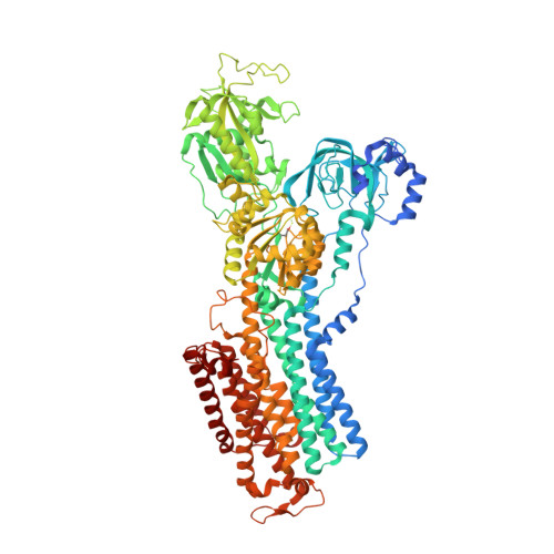
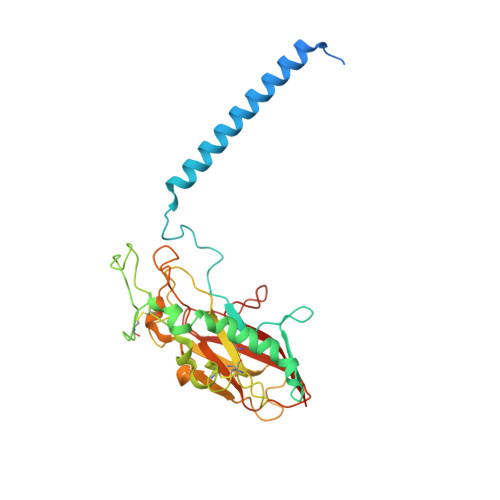
Structural Basis for Binding of Potassium-Competitive Acid Blockers to the Gastric Proton Pump.

Tanaka, S.Morita, M.Yamagishi, T.Madapally, H.V.Hayashida, K.Khandelia, H.Gerle, C.Shigematsu, H.Oshima, A.Abe, K.

(2022) J Med Chem 65: 7843-7853

  • DOI: https://doi.org/10.1021/acs.jmedchem.2c00338
  • Primary Citation Related Structures: 
    7W47, 7W48, 7W49, 7W4A

  • PubMed Abstract: 

    As specific inhibitors of the gastric proton pump, responsible for gastric acidification, K + -competitive acid blockers (P-CABs) have recently been utilized in the clinical treatment of gastric acid-related diseases in Asia. However, as these compounds have been developed based on phenotypic screening, their detailed binding poses are unknown. We show crystal and cryo-EM structures of the gastric proton pump in complex with four different P-CABs, tegoprazan, soraprazan, PF-03716556 and revaprazan, at resolutions reaching 2.8 Å. The structures describe molecular details of their interactions and are supported by functional analyses of mutations and molecular dynamics simulations. We reveal that revaprazan has a novel binding mode in which its tetrahydroisoquinoline moiety binds deep in the cation transport conduit. The mechanism of action of these P-CABs can now be evaluated at the molecular level, which will facilitate the rational development and improvement of currently available P-CABs to provide better treatment of acid-related gastrointestinal diseases.


  • Organizational Affiliation
    • Graduate School of Pharmaceutical Sciences, Nagoya University, Nagoya, 464-8601, Japan.

Macromolecule Content 

  • Total Structure Weight: 149.43 kDa 
  • Atom Count: 9,855 
  • Modeled Residue Count: 1,245 
  • Deposited Residue Count: 1,324 
  • Unique protein chains: 2

Macromolecules

Find similar proteins by:|  3D Structure
Entity ID: 1
MoleculeChains  Sequence LengthOrganismDetailsImage
Potassium-transporting ATPase alpha chain 11,034Sus scrofaMutation(s): 3 
Gene Names: ATP4A
EC: 3.6.3.10 (PDB Primary Data), 7.2.2.19 (UniProt)
Membrane Entity: Yes 
UniProt
Find proteins for P19156 (Sus scrofa)
Explore P19156 
Go to UniProtKB:  P19156
Entity Groups
Sequence Clusters30% Identity50% Identity70% Identity90% Identity95% Identity100% Identity
UniProt GroupP19156
Sequence Annotations
Expand
Reference Sequence
Find similar proteins by:|  3D Structure
Entity ID: 2
MoleculeChains  Sequence LengthOrganismDetailsImage
Potassium-transporting ATPase subunit beta290Sus scrofaMutation(s): 0 
Gene Names: ATP4B
Membrane Entity: Yes 
UniProt
Find proteins for P18434 (Sus scrofa)
Explore P18434 
Go to UniProtKB:  P18434
Entity Groups
Sequence Clusters30% Identity50% Identity70% Identity90% Identity95% Identity100% Identity
UniProt GroupP18434
Glycosylation
Glycosylation Sites: 3
Sequence Annotations
Expand
Reference Sequence

Small Molecules

Ligands 5 Unique
IDChains Name / Formula / InChI Key2D Diagram3D Interactions
PCW

Query on PCW


Download:Ideal Coordinates CCD File
I [auth B]1,2-DIOLEOYL-SN-GLYCERO-3-PHOSPHOCHOLINE
C44 H85 N O8 P
SNKAWJBJQDLSFF-NVKMUCNASA-O
8CK
(Subject of Investigation/LOI)

Query on 8CK


Download:Ideal Coordinates CCD File
C [auth A]N-(4-fluorophenyl)-4,5-dimethyl-6-[(1R)-1-methyl-3,4-dihydro-1H-isoquinolin-2-yl]pyrimidin-2-amine
C22 H23 F N4
LECZXZOBEZITCL-MRXNPFEDSA-N
NAG

Query on NAG


Download:Ideal Coordinates CCD File
F [auth B],
G [auth B],
H [auth B]
2-acetamido-2-deoxy-beta-D-glucopyranose
C8 H15 N O6
OVRNDRQMDRJTHS-FMDGEEDCSA-N
MG

Query on MG


Download:Ideal Coordinates CCD File
E [auth A]MAGNESIUM ION
Mg
JLVVSXFLKOJNIY-UHFFFAOYSA-N
NA

Query on NA


Download:Ideal Coordinates CCD File
D [auth A]SODIUM ION
Na
FKNQFGJONOIPTF-UHFFFAOYSA-N
Modified Residues  1 Unique
IDChains TypeFormula2D DiagramParent
BFD
Query on BFD
A
L-PEPTIDE LINKINGC4 H6 Be F3 N O4ASP

Experimental Data & Validation

Experimental Data

  • Method: ELECTRON MICROSCOPY
  • Resolution: 2.76 Å
  • Aggregation State: PARTICLE 
  • Reconstruction Method: SINGLE PARTICLE 

Structure Validation

View Full Validation Report


Entry History 

& Funding Information

Deposition Data


Funding OrganizationLocationGrant Number
Japan Society for the Promotion of Science (JSPS)Japan21H02426

Revision History  (Full details and data files)

  • Version 1.0: 2022-03-02
    Type: Initial release
  • Version 1.1: 2022-07-20
    Changes: Database references
  • Version 1.2: 2024-10-16
    Changes: Data collection, Structure summary