7W2I | pdb_00007w2i

Crystal structure of LOG (Rv1205) from Mycobacterium tuberculosis


Experimental Data Snapshot

  • Method: X-RAY DIFFRACTION
  • Resolution: 1.80 Å
  • R-Value Free: 
    0.214 (Depositor), 0.223 (DCC) 
  • R-Value Work: 
    0.165 (Depositor), 0.177 (DCC) 
  • R-Value Observed: 
    0.168 (Depositor) 

Starting Model: experimental
View more details

wwPDB Validation 3D Report Full Report

Validation slider image for 7W2I

This is version 1.1 of the entry. See complete history

Literature

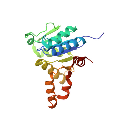
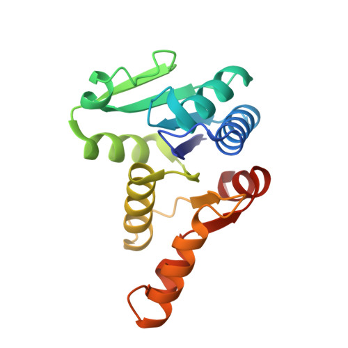
Crystal structure of the cytokinin-producing enzyme "lonely guy" (LOG) from Mycobacterium tuberculosis.

Shang, L.Li, G.Lin, Q.Ou, M.Liang, J.Xiao, G.Wang, Z.Cui, S.Zhang, T.Liu, L.Zhang, G.

(2022) Biochem Biophys Res Commun 598: 113-118

  • DOI: https://doi.org/10.1016/j.bbrc.2022.01.103
  • Primary Citation Related Structures: 
    7W2I

  • PubMed Abstract: 

    Mycobacterium tuberculosis (Mtb) is an extremely successful intracellular pathogen that cause a large number of death worldwide. It is interesting that this non-phytopathogen can synthesize cytokinin by "lonely guy" (LOG) protein. The cytokinin biosynthesis pathway in Mtb is not clear. Here we determined the crystal structure of LOG from Mtb (MtLOG) at a high resolution of 1.8 Å. MtLOG exists as dimer which belongs to type-I LOG and shows a typical α-β Rossmann fold. Like other LOGs, MtLOG also contains a conserved "PGGXGTXXE" motif that contributes to the formation of an active site. For the first time, we found that the MtLOG binds to Mg 2+ in the negative potential pocket. According to the docking result, we found that Arg78, Arg98 and Tyr162 should be the key amino acid involved in substrate binding. Our findings provide a structural basis for cytokinin study in Mtb and will play an important role in design and development of enzyme inhibitors.


  • Organizational Affiliation
    • National Clinical Research Center for Infectious Diseases, Guangdong Provincial Clinical Research Center for Tuberculosis, Shenzhen Third People's Hospital, Southern University of Science and Technology, Shenzhen, 518112, China.

Macromolecule Content 

  • Total Structure Weight: 77.15 kDa 
  • Atom Count: 6,154 
  • Modeled Residue Count: 714 
  • Deposited Residue Count: 714 
  • Unique protein chains: 2

Macromolecules

Find similar proteins by:|  3D Structure
Entity ID: 1
MoleculeChains  Sequence LengthOrganismDetailsImage
Cytokinin riboside 5'-monophosphate phosphoribohydrolase183Mycobacterium tuberculosisMutation(s): 0 
Gene Names: 
EC: 3.2.2
UniProt
Find proteins for O05306 (Mycobacterium tuberculosis (strain ATCC 25618 / H37Rv))
Explore O05306 
Go to UniProtKB:  O05306
Entity Groups
Sequence Clusters30% Identity50% Identity70% Identity90% Identity95% Identity100% Identity
UniProt GroupO05306
Sequence Annotations
Expand
Reference Sequence
Find similar proteins by:|  3D Structure
Entity ID: 2
MoleculeChains  Sequence LengthOrganismDetailsImage
Cytokinin riboside 5'-monophosphate phosphoribohydrolase
B, C, D
177Mycobacterium tuberculosisMutation(s): 0 
Gene Names: 
EC: 3.2.2
UniProt
Find proteins for O05306 (Mycobacterium tuberculosis (strain ATCC 25618 / H37Rv))
Explore O05306 
Go to UniProtKB:  O05306
Entity Groups
Sequence Clusters30% Identity50% Identity70% Identity90% Identity95% Identity100% Identity
UniProt GroupO05306
Sequence Annotations
Expand
Reference Sequence

Small Molecules

Ligands 4 Unique
IDChains Name / Formula / InChI Key2D Diagram3D Interactions
PEG

Query on PEG


Download:Ideal Coordinates CCD File
G [auth A]DI(HYDROXYETHYL)ETHER
C4 H10 O3
MTHSVFCYNBDYFN-UHFFFAOYSA-N
GOL

Query on GOL


Download:Ideal Coordinates CCD File
N [auth D]GLYCEROL
C3 H8 O3
PEDCQBHIVMGVHV-UHFFFAOYSA-N
EDO

Query on EDO


Download:Ideal Coordinates CCD File
F [auth A],
H [auth A],
K [auth C],
M [auth D]
1,2-ETHANEDIOL
C2 H6 O2
LYCAIKOWRPUZTN-UHFFFAOYSA-N
MG

Query on MG


Download:Ideal Coordinates CCD File
E [auth A],
I [auth B],
J [auth C],
L [auth D]
MAGNESIUM ION
Mg
JLVVSXFLKOJNIY-UHFFFAOYSA-N

Experimental Data & Validation

Experimental Data

  • Method: X-RAY DIFFRACTION
  • Resolution: 1.80 Å
  • R-Value Free:  0.214 (Depositor), 0.223 (DCC) 
  • R-Value Work:  0.165 (Depositor), 0.177 (DCC) 
  • R-Value Observed: 0.168 (Depositor) 
Space Group: P 21 21 21
Unit Cell:
Length ( Å )Angle ( ˚ )
a = 67.958α = 90
b = 75.976β = 90
c = 134.953γ = 90
Software Package:
Software NamePurpose
HKL-2000data scaling
REFMACrefinement
PDB_EXTRACTdata extraction
XDSdata reduction
PHASERphasing

Structure Validation

View Full Validation Report


Entry History 

& Funding Information

Deposition Data


Funding OrganizationLocationGrant Number
Other government2019B1515120041

Revision History  (Full details and data files)

  • Version 1.0: 2022-07-06
    Type: Initial release
  • Version 1.1: 2023-11-29
    Changes: Data collection, Refinement description