


OXA-58 crystal structure of acylated meropenem complex 2
Saino, H., Sugiyabu, T., Miyano, M.To be published.
Experimental Data Snapshot
Starting Model: experimental
View more details
Entity ID: 1 | |||||
|---|---|---|---|---|---|
| Molecule | Chains | Sequence Length | Organism | Details | Image |
| Beta-lactamase | 280 | Acinetobacter baumannii | Mutation(s): 0 Gene Names: blaOXA-58, bla-oxa-58, bla-oxa58 EC: 3.5.2.6 | 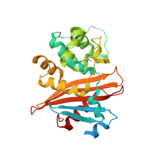 | |
UniProt | |||||
Entity Groups | |||||
| Sequence Clusters | 30% Identity50% Identity70% Identity90% Identity95% Identity100% Identity | ||||
| UniProt Group | Q2TR58 | ||||
Sequence AnnotationsExpand | |||||
Reference Sequence | |||||
| Ligands 3 Unique | |||||
|---|---|---|---|---|---|
| ID | Chains | Name / Formula / InChI Key | 2D Diagram | 3D Interactions | |
| DWZ (Subject of Investigation/LOI) Download:Ideal Coordinates CCD File | D [auth A] | (2S,3R,4S)-4-{[(3S,5S)-5-(dimethylcarbamoyl)pyrrolidin-3-yl]sulfanyl}-2-[(2S,3R)-3-hydroxy-1-oxobutan-2-yl]-3-methyl-3,4-dihydro-2H-pyrrole-5-carboxylic acid C17 H27 N3 O5 S UUIYVKJXUXGPKB-VGWSNGFZSA-N |  | ||
| MER (Subject of Investigation/LOI) Download:Ideal Coordinates CCD File | C [auth A] | (4R,5S)-3-{[(3S,5S)-5-(dimethylcarbamoyl)pyrrolidin-3-yl]sulfanyl}-5-[(2S,3R)-3-hydroxy-1-oxobutan-2-yl]-4-methyl-4,5-d
ihydro-1H-pyrrole-2-carboxylic acid C17 H27 N3 O5 S DYQHXZPAIVAJRU-HTXLXMOSSA-N |  | ||
| SO4 Download:Ideal Coordinates CCD File | B [auth A] | SULFATE ION O4 S QAOWNCQODCNURD-UHFFFAOYSA-L |  | ||
| Modified Residues 1 Unique | |||||
|---|---|---|---|---|---|
| ID | Chains | Type | Formula | 2D Diagram | Parent |
| KCX Query on KCX | A | L-PEPTIDE LINKING | C7 H14 N2 O4 |  | LYS |
| Length ( Å ) | Angle ( ˚ ) |
|---|---|
| a = 75.29 | α = 90 |
| b = 75.29 | β = 90 |
| c = 119.47 | γ = 120 |
| Software Name | Purpose |
|---|---|
| PHENIX | refinement |
| iMOSFLM | data reduction |
| MOLREP | phasing |
| CrystalClear | data collection |
| iMOSFLM | data scaling |
| Funding Organization | Location | Grant Number |
|---|---|---|
| Ministry of Education, Culture, Sports, Science and Technology (Japan) | Japan | S1311005 |
| Ministry of Education, Culture, Sports, Science and Technology (Japan) | Japan | 18K07118 |


