Novel phthalimides regulating PD-1/PD-L1 interaction as potential immunotherapy agents.
Sun, C., Cheng, Y., Liu, X., Wang, G., Min, W., Wang, X., Yuan, K., Hou, Y., Li, J., Zhang, H., Dong, H., Wang, L., Lou, C., Sun, Y., Yu, X., Deng, H., Xiao, Y., Yang, P.(2022) Acta Pharm Sin B 12: 4446-4457
- PubMed: 36561991 Search on PubMedSearch on PubMed Central
- DOI: https://doi.org/10.1016/j.apsb.2022.04.007
- Primary Citation Related Structures:
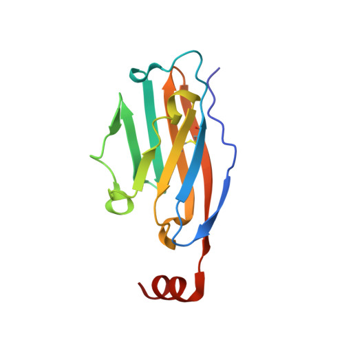
7VUN - PubMed Abstract:
Programmed cell death 1(PD-1)/programmed cell death ligand 1(PD-L1) have emerged as one of the most promising immune checkpoint targets for cancer immunotherapy. Despite the inherent advantages of small-molecule inhibitors over antibodies, the discovery of small-molecule inhibitors has fallen behind that of antibody drugs. Based on docking studies between small molecule inhibitor and PD-L1 protein, changing the chemical linker of inhibitor from a flexible chain to an aromatic ring may improve its binding capacity to PD-L1 protein, which was not reported before. A series of novel phthalimide derivatives from structure-based rational design was synthesized. P39 was identified as the best inhibitor with promising activity, which not only inhibited PD-1/PD-L1 interaction (IC 50 = 8.9 nmol/L), but also enhanced killing efficacy of immune cells on cancer cells. Co-crystal data demonstrated that P39 induced the dimerization of PD-L1 proteins, thereby blocking the binding of PD-1/PD-L1. Moreover, P39 exhibited a favorable safety profile with a LD 50 > 5000 mg/kg and showed significant in vivo antitumor activity through promoting CD8 + T cell activation. All these data suggest that P39 acts as a promising small chemical inhibitor against the PD-1/PD-L1 axis and has the potential to improve the immunotherapy efficacy of T-cells.
- State Key Laboratory of Natural Medicines and Jiangsu Key Laboratory of Drug Design and Optimization, China Pharmaceutical University, Nanjing 210009, China.
Organizational Affiliation: