7VR7 | pdb_00007vr7

Inward-facing structure of human EAAT2 in the WAY213613-bound state


Experimental Data Snapshot

  • Method: ELECTRON MICROSCOPY
  • Resolution: 3.49 Å
  • Aggregation State: PARTICLE 
  • Reconstruction Method: SINGLE PARTICLE 

wwPDB Validation 3D Report Full Report

Validation slider image for 7VR7

This is version 1.2 of the entry. See complete history

Literature

Structural insights into inhibitory mechanism of human excitatory amino acid transporter EAAT2.

Kato, T.Kusakizako, T.Jin, C.Zhou, X.Ohgaki, R.Quan, L.Xu, M.Okuda, S.Kobayashi, K.Yamashita, K.Nishizawa, T.Kanai, Y.Nureki, O.

(2022) Nat Commun 13: 4714-4714

  • DOI: https://doi.org/10.1038/s41467-022-32442-6
  • Primary Citation Related Structures: 
    7VR7, 7VR8

  • PubMed Abstract: 

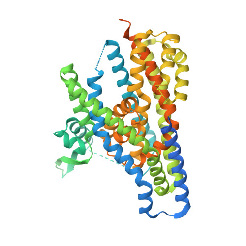
    Glutamate is a pivotal excitatory neurotransmitter in mammalian brains, but excessive glutamate causes numerous neural disorders. Almost all extracellular glutamate is retrieved by the glial transporter, Excitatory Amino Acid Transporter 2 (EAAT2), belonging to the SLC1A family. However, in some cancers, EAAT2 expression is enhanced and causes resistance to therapies by metabolic disturbance. Despite its crucial roles, the detailed structural information about EAAT2 has not been available. Here, we report cryo-EM structures of human EAAT2 in substrate-free and selective inhibitor WAY213613-bound states at 3.2 Å and 2.8 Å, respectively. EAAT2 forms a trimer, with each protomer consisting of transport and scaffold domains. Along with a glutamate-binding site, the transport domain possesses a cavity that could be disrupted during the transport cycle. WAY213613 occupies both the glutamate-binding site and cavity of EAAT2 to interfere with its alternating access, where the sensitivity is defined by the inner environment of the cavity. We provide the characterization of the molecular features of EAAT2 and its selective inhibition mechanism that may facilitate structure-based drug design for EAAT2.


  • Organizational Affiliation
    • Department of Biological Science, Graduate School of Science, The University of Tokyo, Tokyo, Japan.

Macromolecule Content 

  • Total Structure Weight: 66.33 kDa 
  • Atom Count: 3,187 
  • Modeled Residue Count: 402 
  • Deposited Residue Count: 581 
  • Unique protein chains: 1

Macromolecules

Find similar proteins by:|  3D Structure
Entity ID: 1
MoleculeChains  Sequence LengthOrganismDetailsImage
Excitatory amino acid transporter 2581Homo sapiensMutation(s): 0 
Gene Names: SLC1A2EAAT2GLT1
Membrane Entity: Yes 
UniProt & NIH Common Fund Data Resources
Find proteins for P43004 (Homo sapiens)
Explore P43004 
Go to UniProtKB:  P43004
PHAROS:  P43004
GTEx:  ENSG00000110436 
Entity Groups
Sequence Clusters30% Identity50% Identity70% Identity90% Identity95% Identity100% Identity
UniProt GroupP43004
Sequence Annotations
Expand
Reference Sequence

Small Molecules

Ligands 4 Unique
IDChains Name / Formula / InChI Key2D Diagram3D Interactions
PC1

Query on PC1


Download:Ideal Coordinates CCD File
F [auth A],
G [auth A]
1,2-DIACYL-SN-GLYCERO-3-PHOSPHOCHOLINE
C44 H88 N O8 P
NRJAVPSFFCBXDT-HUESYALOSA-N
9Z9

Query on 9Z9


Download:Ideal Coordinates CCD File
C [auth A](3beta,14beta,17beta,25R)-3-[4-methoxy-3-(methoxymethyl)butoxy]spirost-5-en
C34 H56 O5
CEEBZAXXSRFQIC-GZSGZGDASA-N
GJ0
(Subject of Investigation/LOI)

Query on GJ0


Download:Ideal Coordinates CCD File
B [auth A](2S)-2-azanyl-4-[[4-[2-bromanyl-4,5-bis(fluoranyl)phenoxy]phenyl]amino]-4-oxidanylidene-butanoic acid
C16 H13 Br F2 N2 O4
BNYDDAAZMBUFRG-ZDUSSCGKSA-N
CLR
(Subject of Investigation/LOI)

Query on CLR


Download:Ideal Coordinates CCD File
D [auth A],
E [auth A]
CHOLESTEROL
C27 H46 O
HVYWMOMLDIMFJA-DPAQBDIFSA-N
Binding Affinity Annotations 
IDSourceBinding Affinity
GJ0 BindingDB:  7VR7 Ki: 85 (nM) from 1 assay(s)
IC50: min: 71, max: 130 (nM) from 3 assay(s)

Experimental Data & Validation

Experimental Data

  • Method: ELECTRON MICROSCOPY
  • Resolution: 3.49 Å
  • Aggregation State: PARTICLE 
  • Reconstruction Method: SINGLE PARTICLE 
EM Software:
TaskSoftware PackageVersion
MODEL REFINEMENTREFMAC5.8.0291
RECONSTRUCTIONRELION3.1

Structure Validation

View Full Validation Report


Entry History 

& Funding Information

Deposition Data


Funding OrganizationLocationGrant Number
Japan Society for the Promotion of Science (JSPS)Japan--

Revision History  (Full details and data files)

  • Version 1.0: 2022-08-10
    Type: Initial release
  • Version 1.1: 2022-09-28
    Changes: Database references
  • Version 1.2: 2024-06-26
    Changes: Data collection, Refinement description