


Distinct structural characteristics define a new subfamily of Mycoplasma ferritin
Wang, W., Liu, X., Wang, Y., Wang, Y., Fu, D., Xi, H., Zhao, Y., Wang, H.(2022) Chin Chem Lett 33: 4952-4955
Experimental Data Snapshot
Starting Model: in silico
View more details
wwPDB Validation 3D Report Full Report

Entity ID: 1 | |||||
|---|---|---|---|---|---|
| Molecule | Chains | Sequence Length | Organism | Details | Image |
| Ferritin-like diiron domain-containing protein | 172 | Ureaplasma urealyticum | Mutation(s): 0 Gene Names: EPH05_02360 | 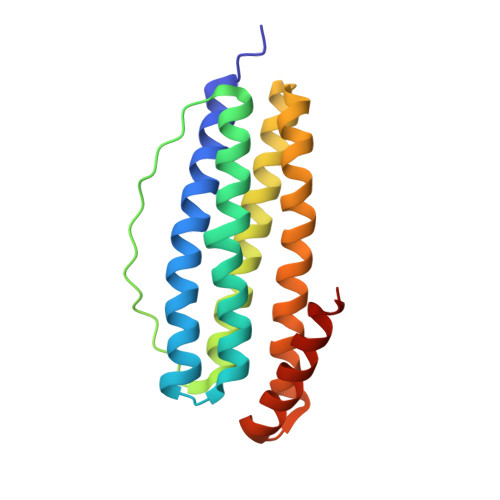 | |
| Ligands 2 Unique | |||||
|---|---|---|---|---|---|
| ID | Chains | Name / Formula / InChI Key | 2D Diagram | 3D Interactions | |
| FE Download:Ideal Coordinates CCD File | B [auth A], C [auth A], D [auth A], E [auth A] | FE (III) ION Fe VTLYFUHAOXGGBS-UHFFFAOYSA-N |  | ||
| CL (Subject of Investigation/LOI) Download:Ideal Coordinates CCD File | F [auth A], G [auth A], H [auth A] | CHLORIDE ION Cl VEXZGXHMUGYJMC-UHFFFAOYSA-M |  | ||
| Length ( Å ) | Angle ( ˚ ) |
|---|---|
| a = 178.441 | α = 90 |
| b = 178.441 | β = 90 |
| c = 178.441 | γ = 90 |
| Software Name | Purpose |
|---|---|
| XSCALE | data scaling |
| PHASER | phasing |
| PHENIX | refinement |
| PDB_EXTRACT | data extraction |
| HKL-2000 | data reduction |
| Funding Organization | Location | Grant Number |
|---|---|---|
| National Natural Science Foundation of China (NSFC) | China | 62075118, 21601112 |


