7VNN

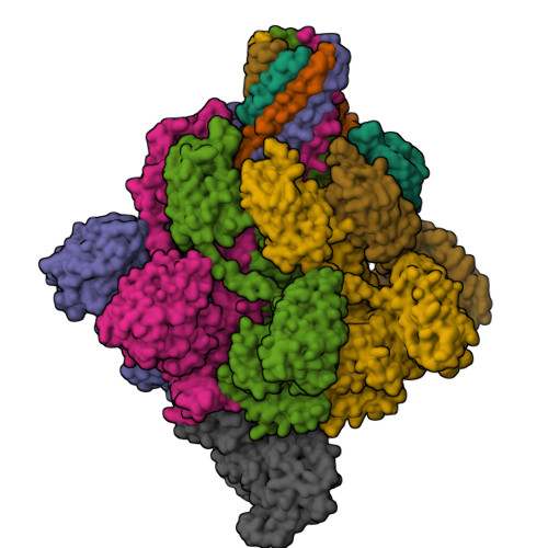
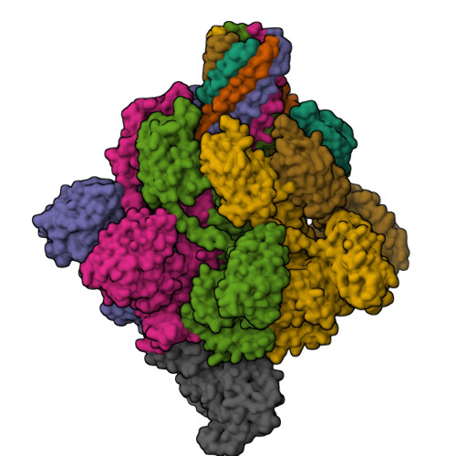
Complex structure of Clostridioides difficile enzymatic component (CDTa) and binding component (CDTb) pore with long stem


Experimental Data Snapshot

  • Method: ELECTRON MICROSCOPY
  • Resolution: 2.64 Å
  • Aggregation State: PARTICLE 
  • Reconstruction Method: SINGLE PARTICLE 

Starting Models: experimental
View more details

wwPDB Validation   3D Report Full Report


This is version 1.2 of the entry. See complete history


Literature

Cryo-EM structures of the translocational binary toxin complex CDTa-bound CDTb-pore from Clostridioides difficile.

Kawamoto, A.Yamada, T.Yoshida, T.Sato, Y.Kato, T.Tsuge, H.

(2022) Nat Commun 13: 6119-6119

  • DOI: https://doi.org/10.1038/s41467-022-33888-4
  • Primary Citation of Related Structures:  
    7VNJ, 7VNN, 7YVQ, 7YVS

  • PubMed Abstract: 

    Some bacteria express a binary toxin translocation system, consisting of an enzymatic subunit and translocation pore, that delivers enzymes into host cells through endocytosis. The most clinically important bacterium with such a system is Clostridioides difficile (formerly Clostridium). The CDTa and CDTb proteins from its system represent important therapeutic targets. CDTb has been proposed to be a di-heptamer, but its physiological heptameric structure has not yet been reported. Here, we report the cryo-EM structure of CDTa bound to the CDTb-pore, which reveals that CDTa binding induces partial unfolding and tilting of the first CDTa α-helix. In the CDTb-pore, an NSS-loop exists in 'in' and 'out' conformations, suggesting its involvement in substrate translocation. Finally, 3D variability analysis revealed CDTa movements from a folded to an unfolded state. These dynamic structural information provide insights into drug design against hypervirulent C. difficile strains.


  • Organizational Affiliation

    Institute for Protein Research, Osaka University, Suita, Osaka, 565-0871, Japan.


Macromolecules
Find similar proteins by:  (by identity cutoff)  |  3D Structure
Entity ID: 1
MoleculeChains Sequence LengthOrganismDetailsImage
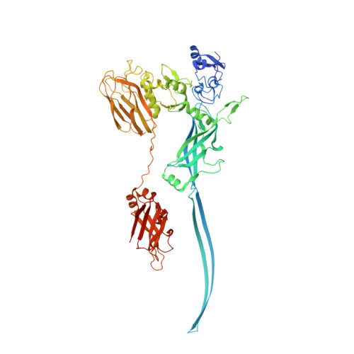
ADP-ribosylating binary toxin binding subunit CdtB
A, B, C, D, E
A, B, C, D, E, F, G
675Clostridioides difficileMutation(s): 0 
Gene Names: cdtB
Membrane Entity: Yes 
UniProt
Find proteins for A8DS70 (Clostridioides difficile)
Explore A8DS70 
Go to UniProtKB:  A8DS70
Entity Groups  
Sequence Clusters30% Identity50% Identity70% Identity90% Identity95% Identity100% Identity
UniProt GroupA8DS70
Sequence Annotations
Expand
  • Reference Sequence
Find similar proteins by:  (by identity cutoff)  |  3D Structure
Entity ID: 2
MoleculeChains Sequence LengthOrganismDetailsImage
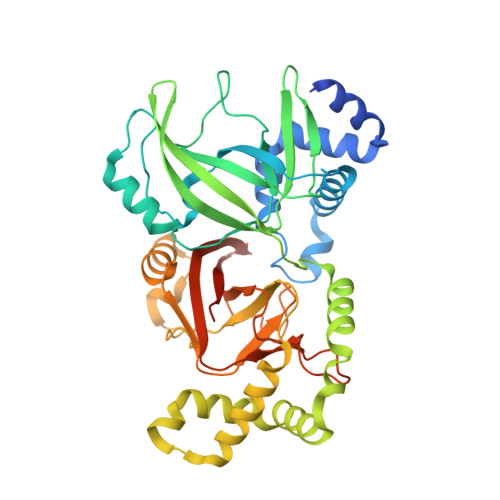
CdtA428Clostridioides difficileMutation(s): 0 
Gene Names: cdtA
UniProt
Find proteins for Q9KH42 (Clostridioides difficile)
Explore Q9KH42 
Go to UniProtKB:  Q9KH42
Entity Groups  
Sequence Clusters30% Identity50% Identity70% Identity90% Identity95% Identity100% Identity
UniProt GroupQ9KH42
Sequence Annotations
Expand
  • Reference Sequence
Small Molecules
Ligands 1 Unique
IDChains Name / Formula / InChI Key2D Diagram3D Interactions
CA (Subject of Investigation/LOI)
Query on CA

Download Ideal Coordinates CCD File 
AA [auth G]
BA [auth G]
CA [auth G]
I [auth A]
J [auth A]
AA [auth G],
BA [auth G],
CA [auth G],
I [auth A],
J [auth A],
K [auth A],
L [auth B],
M [auth B],
N [auth B],
O [auth C],
P [auth C],
Q [auth C],
R [auth D],
S [auth D],
T [auth D],
U [auth E],
V [auth E],
W [auth E],
X [auth F],
Y [auth F],
Z [auth F]
CALCIUM ION
Ca
BHPQYMZQTOCNFJ-UHFFFAOYSA-N
Experimental Data & Validation

Experimental Data

  • Method: ELECTRON MICROSCOPY
  • Resolution: 2.64 Å
  • Aggregation State: PARTICLE 
  • Reconstruction Method: SINGLE PARTICLE 
EM Software:
TaskSoftware PackageVersion
MODEL REFINEMENTPHENIX
MODEL REFINEMENTCoot
RECONSTRUCTIONRELION3.1

Structure Validation

View Full Validation Report


Entry History & Funding Information

Deposition Data


Funding OrganizationLocationGrant Number
Japan Society for the Promotion of Science (JSPS)Japan--
Japan Science and TechnologyJapan--

Revision History  (Full details and data files)

  • Version 1.0: 2022-10-26
    Type: Initial release
  • Version 1.1: 2022-11-02
    Changes: Database references
  • Version 1.2: 2024-06-19
    Changes: Data collection, Refinement description