7VN9 | pdb_00007vn9

Crystal structure of human coronavirus 229E spike protein receptor-binding domain in complex with C04 Fab


Experimental Data Snapshot

  • Method: X-RAY DIFFRACTION
  • Resolution: 4.49 Å
  • R-Value Free: 
    0.288 (Depositor), 0.306 (DCC) 
  • R-Value Work: 
    0.273 (Depositor), 0.288 (DCC) 
  • R-Value Observed: 
    0.274 (Depositor) 

Starting Models: experimental
View more details

wwPDB Validation 3D Report Full Report

Validation slider image for 7VN9

Ligand Structure Quality Assessment 


This is version 1.3 of the entry. See complete history

Literature

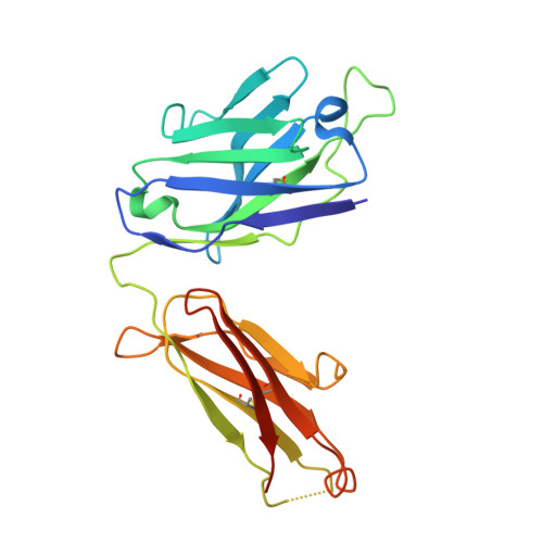
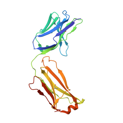
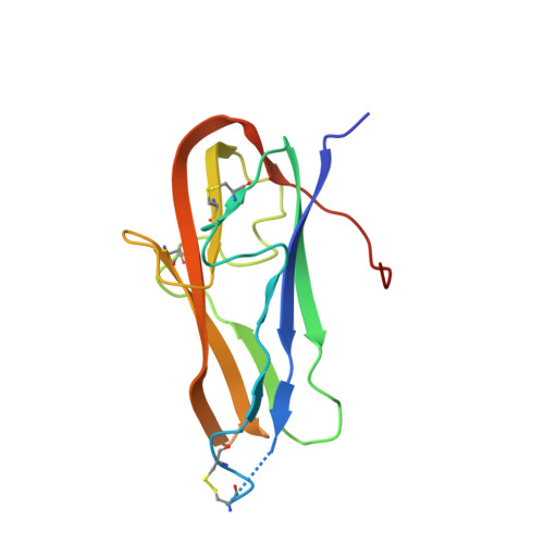
Antigenic mapping reveals sites of vulnerability on alpha-HCoV spike protein.

Xiang, J.Su, J.Lan, Q.Zhao, W.Zhou, Y.Xu, Y.Niu, J.Xia, S.Qi, Q.Sidhu, S.Lu, L.Miersch, S.Yang, B.

(2022) Commun Biol 5: 1179-1179

  • DOI: https://doi.org/10.1038/s42003-022-04160-8
  • Primary Citation Related Structures: 
    7VMZ, 7VN9, 7VNG

  • PubMed Abstract: 

    Understanding the antigenic signatures of all human coronaviruses (HCoVs) Spike (S) proteins is imperative for pan-HCoV epitopes identification and broadly effective vaccine development. To depict the currently elusive antigenic signatures of α-HCoVs S proteins, we isolated a panel of antibodies against the HCoV-229E S protein and characterized their epitopes and neutralizing potential. We found that the N-terminal domain of HCoV-229E S protein is antigenically dominant wherein an antigenic supersite is present and appears conserved in HCoV-NL63, which holds potential to serve as a pan-α-HCoVs epitope. In the receptor binding domain, a neutralizing epitope is captured in the end distal to the receptor binding site, reminiscent of the locations of the SARS-CoV-2 RBD cryptic epitopes. We also identified a neutralizing antibody that recognizes the connector domain, thus representing the first S2-directed neutralizing antibody against α-HCoVs. The unraveled HCoVs S proteins antigenic similarities and variances among genera highlight the challenges faced by pan-HCoV vaccine design while supporting the feasibility of broadly effective vaccine development against a subset of HCoVs.


  • Organizational Affiliation
    • School of Life Science and Technology and Shanghai Institute for Advanced Immunochemical Studies, ShanghaiTech University, 393 Middle Huaxia Road, 201210, Shanghai, China.

Macromolecule Content 

  • Total Structure Weight: 129.5 kDa 
  • Atom Count: 8,541 
  • Modeled Residue Count: 1,113 
  • Deposited Residue Count: 1,188 
  • Unique protein chains: 3

Macromolecules

Find similar proteins by:|  3D Structure
Entity ID: 1
MoleculeChains  Sequence LengthOrganismDetailsImage
C04 Fab heavy chainA [auth H],
D [auth A]
231Homo sapiensMutation(s): 0 
Find similar proteins by:|  3D Structure
Entity ID: 2
MoleculeChains  Sequence LengthOrganismDetailsImage
C04 Fab light chainB [auth L],
E [auth B]
215Homo sapiensMutation(s): 0 
Find similar proteins by:|  3D Structure
Entity ID: 3
MoleculeChains  Sequence LengthOrganismDetailsImage
Spike glycoprotein S1C [auth E],
F [auth C]
148Human coronavirus 229EMutation(s): 0 
Gene Names: S2
UniProt
Find proteins for P15423 (Human coronavirus 229E)
Explore P15423 
Go to UniProtKB:  P15423
Entity Groups
Sequence Clusters30% Identity50% Identity70% Identity90% Identity95% Identity100% Identity
UniProt GroupP15423
Glycosylation
Glycosylation Sites: 2
Sequence Annotations
Expand
Reference Sequence

Oligosaccharides

Help  
Entity ID: 4
MoleculeChains Length2D Diagram GlycosylationD Interactions
beta-D-mannopyranose-(1-4)-2-acetamido-2-deoxy-beta-D-glucopyranose-(1-4)-2-acetamido-2-deoxy-beta-D-glucopyranoseG [auth D],
I [auth G]
3N-Glycosylation
Glycosylation Resources
GlyTouCan: G15407YE
GlyCosmos: G15407YE
GlyGen: G15407YE
Entity ID: 5
MoleculeChains Length2D Diagram GlycosylationD Interactions
2-acetamido-2-deoxy-beta-D-glucopyranose-(1-4)-2-acetamido-2-deoxy-beta-D-glucopyranoseH [auth F]2N-Glycosylation
Glycosylation Resources
GlyTouCan: G42666HT
GlyCosmos: G42666HT
GlyGen: G42666HT

Small Molecules

Ligands 1 Unique
IDChains Name / Formula / InChI Key2D Diagram3D Interactions
NAG

Query on NAG


Download:Ideal Coordinates CCD File
J [auth C]2-acetamido-2-deoxy-beta-D-glucopyranose
C8 H15 N O6
OVRNDRQMDRJTHS-FMDGEEDCSA-N

Experimental Data & Validation

Experimental Data

  • Method: X-RAY DIFFRACTION
  • Resolution: 4.49 Å
  • R-Value Free:  0.288 (Depositor), 0.306 (DCC) 
  • R-Value Work:  0.273 (Depositor), 0.288 (DCC) 
  • R-Value Observed: 0.274 (Depositor) 
Space Group: P 62 2 2
Unit Cell:
Length ( Å )Angle ( ˚ )
a = 183.732α = 90
b = 183.732β = 90
c = 255.988γ = 120
Software Package:
Software NamePurpose
PHENIXrefinement
PDB_EXTRACTdata extraction
HKL-2000data reduction
HKL-2000data scaling
PHASERphasing

Structure Validation

View Full Validation Report


Ligand Structure Quality Assessment 


Entry History 

& Funding Information

Deposition Data


Funding OrganizationLocationGrant Number
National Science Foundation (NSF, China)China32070170

Revision History  (Full details and data files)

  • Version 1.0: 2022-10-26
    Type: Initial release
  • Version 1.1: 2022-11-23
    Changes: Database references
  • Version 1.2: 2023-11-29
    Changes: Data collection, Refinement description
  • Version 1.3: 2024-10-16
    Changes: Structure summary