Structure Characterization of Escherichia coli Pseudouridine Kinase PsuK.
Li, X., Li, K., Guo, W., Wen, Y., Meng, C., Wu, B.(2022) Front Microbiol 13: 926099-926099
- PubMed: 35783380 Search on PubMedSearch on PubMed Central
- DOI: https://doi.org/10.3389/fmicb.2022.926099
- Primary Citation Related Structures:
7VKP, 7VSK, 7W93 - PubMed Abstract:
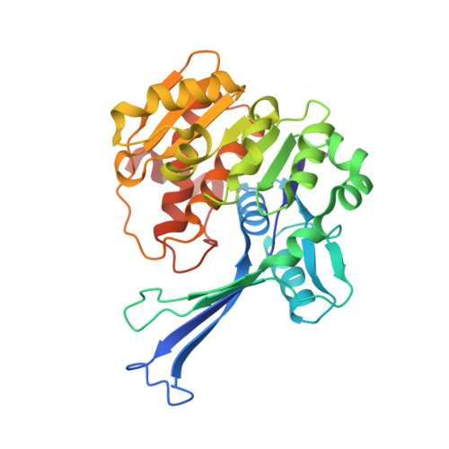
Pseudouridine (Ψ) is one of the most abundant RNA modifications in cellular RNAs that post-transcriptionally impact many aspects of RNA. However, the metabolic fate of modified RNA nucleotides has long been a question. A pseudouridine kinase (PsuK) and a pseudouridine monophosphate glycosylase (PsuG) in Escherichia coli were first characterized as involved in pseudouridine degradation by catalyzing the phosphorylation of pseudouridine to pseudouridine 5'-phosphate (ΨMP) and further hydrolyzing 5'-ΨMP to produce uracil and ribose 5'-phosphate. Recently, their homolog proteins in eukaryotes were also identified, which were named PUKI and PUMY in Arabidopsis . Here, we solved the crystal structures of apo- Ec PsuK and its binary complex with Ψ or N 1 -methyl-pseudouridine (m1Ψ). The structure of Ec PsuK showed a homodimer conformation assembled by its β-thumb region. Ec PsuK has an appropriate binding site with a series of hydrophilic and hydrophobic interactions for Ψ. Moreover, our complex structure of Ec PsuK-m1Ψ suggested the binding pocket has an appropriate capacity for m1Ψ. We also identified the monovalent ion-binding site and potential ATP-binding site. Our studies improved the understanding of the mechanism of Ψ turnover.
- Guangdong Provincial Key Laboratory of Malignant Tumor Epigenetics and Gene Regulation, Guangdong-Hong Kong Joint Laboratory for RNA Medicine, RNA Biomedical Institute, Medical Research Center, Sun Yat-sen Memorial Hospital, Sun Yat-sen University, Guangzhou, China.
Organizational Affiliation: