7VH5

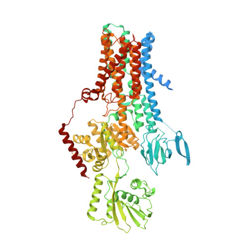
Cryo-EM structure of the hexameric plasma membrane H+-ATPase in the autoinhibited state (pH 7.4, C1 symmetry)


Experimental Data Snapshot

  • Method: ELECTRON MICROSCOPY
  • Resolution: 3.20 Å
  • Aggregation State: PARTICLE 
  • Reconstruction Method: SINGLE PARTICLE 

wwPDB Validation   3D Report Full Report


This is version 1.2 of the entry. See complete history


Literature

Structure and activation mechanism of the hexameric plasma membrane H + -ATPase.

Zhao, P.Zhao, C.Chen, D.Yun, C.Li, H.Bai, L.

(2021) Nat Commun 12: 6439-6439

  • DOI: https://doi.org/10.1038/s41467-021-26782-y
  • Primary Citation of Related Structures:  
    7VH5, 7VH6

  • PubMed Abstract: 

    The S. cerevisiae plasma membrane H + -ATPase, Pma1, is a P3A-type ATPase and the primary protein component of the membrane compartment of Pma1 (MCP). Like other plasma membrane H + -ATPases, Pma1 assembles and functions as a hexamer, a property unique to this subfamily among the larger family of P-type ATPases. It has been unclear how Pma1 organizes the yeast membrane into MCP microdomains, or why it is that Pma1 needs to assemble into a hexamer to establish the membrane electrochemical proton gradient. Here we report a high-resolution cryo-EM study of native Pma1 hexamers embedded in endogenous lipids. Remarkably, we found that the Pma1 hexamer encircles a liquid-crystalline membrane domain composed of 57 ordered lipid molecules. The Pma1-encircled lipid patch structure likely serves as the building block of the MCP. At pH 7.4, the carboxyl-terminal regulatory α-helix binds to the phosphorylation domains of two neighboring Pma1 subunits, locking the hexamer in the autoinhibited state. The regulatory helix becomes disordered at lower pH, leading to activation of the Pma1 hexamer. The activation process is accompanied by a 6.7 Å downward shift and a 40° rotation of transmembrane helices 1 and 2 that line the proton translocation path. The conformational changes have enabled us to propose a detailed mechanism for ATP-hydrolysis-driven proton pumping across the plasma membrane. Our structures will facilitate the development of antifungal drugs that target this essential protein.


  • Organizational Affiliation

    Department of Biochemistry and Biophysics, School of Basic Medical Sciences, Peking University, Beijing, China.


Macromolecules
Find similar proteins by:  (by identity cutoff)  |  3D Structure
Entity ID: 1
MoleculeChains Sequence LengthOrganismDetailsImage
Plasma membrane ATPase 1
A, B, C, D, E
A, B, C, D, E, F
918Saccharomyces cerevisiae S288CMutation(s): 0 
Gene Names: PMA1YGL008C
EC: 7.1.2.1
Membrane Entity: Yes 
UniProt
Find proteins for P05030 (Saccharomyces cerevisiae (strain ATCC 204508 / S288c))
Explore P05030 
Go to UniProtKB:  P05030
Entity Groups  
Sequence Clusters30% Identity50% Identity70% Identity90% Identity95% Identity100% Identity
UniProt GroupP05030
Sequence Annotations
Expand
  • Reference Sequence
Small Molecules
Ligands 2 Unique
IDChains Name / Formula / InChI Key2D Diagram3D Interactions
POV
Query on POV

Download Ideal Coordinates CCD File 
AA [auth C]
AB [auth F]
BA [auth C]
BB [auth F]
CA [auth C]
AA [auth C],
AB [auth F],
BA [auth C],
BB [auth F],
CA [auth C],
CB [auth F],
DA [auth C],
DB [auth F],
EA [auth C],
EB [auth F],
FA [auth D],
G [auth A],
GA [auth D],
H [auth A],
HA [auth D],
I [auth A],
IA [auth D],
J [auth A],
JA [auth D],
K [auth A],
KA [auth D],
L [auth A],
LA [auth D],
M [auth A],
MA [auth D],
N [auth A],
NA [auth E],
O [auth B],
OA [auth E],
P [auth B],
PA [auth E],
Q [auth B],
QA [auth E],
R [auth B],
RA [auth E],
S [auth B],
SA [auth E],
T [auth B],
TA [auth E],
U [auth B],
UA [auth E],
V [auth B],
VA [auth E],
W [auth C],
WA [auth F],
X [auth C],
XA [auth F],
Y [auth C],
YA [auth F],
Z [auth C],
ZA [auth F]
(2S)-3-(hexadecanoyloxy)-2-[(9Z)-octadec-9-enoyloxy]propyl 2-(trimethylammonio)ethyl phosphate
C42 H82 N O8 P
WTJKGGKOPKCXLL-PFDVCBLKSA-N
SPH
Query on SPH

Download Ideal Coordinates CCD File 
FB [auth F]SPHINGOSINE
C18 H37 N O2
WWUZIQQURGPMPG-MSOLQXFVSA-N
Experimental Data & Validation

Experimental Data

  • Method: ELECTRON MICROSCOPY
  • Resolution: 3.20 Å
  • Aggregation State: PARTICLE 
  • Reconstruction Method: SINGLE PARTICLE 

Structure Validation

View Full Validation Report


Entry History & Funding Information

Deposition Data


Funding OrganizationLocationGrant Number
National Natural Science Foundation of China (NSFC)China32171212

Revision History  (Full details and data files)

  • Version 1.0: 2021-11-24
    Type: Initial release
  • Version 1.1: 2022-02-16
    Changes: Database references
  • Version 1.2: 2024-06-19
    Changes: Advisory, Data collection, Derived calculations