7VDY | pdb_00007vdy

Crystal structure of O-ureidoserine racemase


Experimental Data Snapshot

  • Method: X-RAY DIFFRACTION
  • Resolution: 2.12 Å
  • R-Value Free: 
    0.210 (Depositor), 0.210 (DCC) 
  • R-Value Work: 
    0.166 (Depositor), 0.167 (DCC) 
  • R-Value Observed: 
    0.168 (Depositor) 

Starting Model: experimental
View more details

wwPDB Validation 3D Report Full Report

Validation slider image for 7VDY

This is version 1.2 of the entry. See complete history

Literature

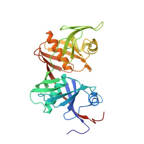
Crystal structure of O-ureidoserine racemase found in the d-cycloserine biosynthetic pathway.

Oda, K.Sakaguchi, T.Matoba, Y.

(2022) Proteins 90: 912-918

  • DOI: https://doi.org/10.1002/prot.26290
  • Primary Citation Related Structures: 
    7VDY

  • PubMed Abstract: 

    The O-ureidoserine racemase (DcsC) is an enzyme found from the biosynthetic gene cluster of antitubercular agent d-cycloserine. Although DcsC is homologous to diaminopimelate epimerase (DapF) that catalyzes the interconversion between ll- and dl-diaminopimelic acid, it specifically catalyzes the interconversion between O-ureido-l-serine and its enantiomer. Here we determined the crystal structure of DcsC at a resolution of 2.12 Å, implicating that the catalytic mechanism of DcsC shares similarity with that of DapF. Comparing the structure of the active center of DcsC to that of DapF, Thr72, Thr198, and Tyr219 of DcsC are likely to be involved in the substrate specificity.


  • Organizational Affiliation
    • Department of Virology, Institute of Biomedical and Health Sciences, Hiroshima University, Hiroshima, Japan.

Macromolecule Content 

  • Total Structure Weight: 63.94 kDa 
  • Atom Count: 4,654 
  • Modeled Residue Count: 564 
  • Deposited Residue Count: 590 
  • Unique protein chains: 1

Macromolecules

Find similar proteins by:|  3D Structure
Entity ID: 1
MoleculeChains  Sequence LengthOrganismDetailsImage
O-ureido-serine racemase
A, B
295Streptomyces lavendulaeMutation(s): 0 
Gene Names: dcsCdapF
EC: 5.1.1.19
UniProt
Find proteins for D2Z026 (Streptomyces lavendulae)
Explore D2Z026 
Go to UniProtKB:  D2Z026
Entity Groups
Sequence Clusters30% Identity50% Identity70% Identity90% Identity95% Identity100% Identity
UniProt GroupD2Z026
Sequence Annotations
Expand
Reference Sequence

Small Molecules

Ligands 1 Unique
IDChains Name / Formula / InChI Key2D Diagram3D Interactions
SO4

Query on SO4


Download:Ideal Coordinates CCD File
C [auth A]
D [auth A]
E [auth A]
F [auth A]
G [auth A]
C [auth A],
D [auth A],
E [auth A],
F [auth A],
G [auth A],
H [auth A],
I [auth B],
J [auth B],
K [auth B],
L [auth B]
SULFATE ION
O4 S
QAOWNCQODCNURD-UHFFFAOYSA-L

Experimental Data & Validation

Experimental Data

  • Method: X-RAY DIFFRACTION
  • Resolution: 2.12 Å
  • R-Value Free:  0.210 (Depositor), 0.210 (DCC) 
  • R-Value Work:  0.166 (Depositor), 0.167 (DCC) 
  • R-Value Observed: 0.168 (Depositor) 
Space Group: P 1 21 1
Unit Cell:
Length ( Å )Angle ( ˚ )
a = 53.121α = 90
b = 96.051β = 92.13
c = 57.286γ = 90
Software Package:
Software NamePurpose
PHENIXrefinement
Aimlessdata scaling
PDB_EXTRACTdata extraction
XDSdata reduction
PHASERphasing

Structure Validation

View Full Validation Report


Entry History 

& Funding Information

Deposition Data


Funding OrganizationLocationGrant Number
Not funded--

Revision History  (Full details and data files)

  • Version 1.0: 2021-12-15
    Type: Initial release
  • Version 1.1: 2022-03-16
    Changes: Database references
  • Version 1.2: 2023-11-29
    Changes: Data collection, Refinement description