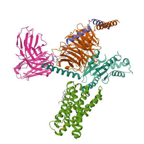
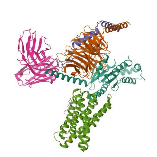
7VDL

Cryo-EM structure of pseudoallergen receptor MRGPRX2 complex with circular cortistatin-14


Experimental Data Snapshot

  • Method: ELECTRON MICROSCOPY
  • Resolution: 3.22 Å
  • Aggregation State: PARTICLE 
  • Reconstruction Method: SINGLE PARTICLE 

wwPDB Validation   3D Report Full Report


This is version 1.2 of the entry. See complete history


Literature

Structure, function and pharmacology of human itch receptor complexes.

Yang, F.Guo, L.Li, Y.Wang, G.Wang, J.Zhang, C.Fang, G.X.Chen, X.Liu, L.Yan, X.Liu, Q.Qu, C.Xu, Y.Xiao, P.Zhu, Z.Li, Z.Zhou, J.Yu, X.Gao, N.Sun, J.P.

(2021) Nature 600: 164-169

  • DOI: https://doi.org/10.1038/s41586-021-04077-y
  • Primary Citation of Related Structures:  
    7VDH, 7VDL, 7VDM, 7VUY, 7VUZ, 7VV0, 7VV3, 7VV4, 7VV5, 7VV6

  • PubMed Abstract: 

    In the clades of animals that diverged from the bony fish, a group of Mas-related G-protein-coupled receptors (MRGPRs) evolved that have an active role in itch and allergic signals 1,2 . As an MRGPR, MRGPRX2 is known to sense basic secretagogues (agents that promote secretion) and is involved in itch signals and eliciting pseudoallergic reactions 3-6 . MRGPRX2 has been targeted by drug development efforts to prevent the side effects induced by certain drugs or to treat allergic diseases. Here we report a set of cryo-electron microscopy structures of the MRGPRX2-G i1 trimer in complex with polycationic compound 48/80 or with inflammatory peptides. The structures of the MRGPRX2-G i1 complex exhibited shallow, solvent-exposed ligand-binding pockets. We identified key common structural features of MRGPRX2 and describe a consensus motif for peptidic allergens. Beneath the ligand-binding pocket, the unusual kink formation at transmembrane domain 6 (TM6) and the replacement of the general toggle switch from Trp 6.48 to Gly 6.48 (superscript annotations as per Ballesteros-Weinstein nomenclature) suggest a distinct activation process. We characterized the interfaces of MRGPRX2 and the G i trimer, and mapped the residues associated with key single-nucleotide polymorphisms on both the ligand and G-protein interfaces of MRGPRX2. Collectively, our results provide a structural basis for the sensing of cationic allergens by MRGPRX2, potentially facilitating the rational design of therapies to prevent unwanted pseudoallergic reactions.


  • Organizational Affiliation

    Key Laboratory of Molecular Cardiovascular Science, Department of Physiology and Pathophysiology, School of Basic Medical Sciences, Peking University, Ministry of Education, Beijing, China.


Macromolecules
Find similar proteins by:  (by identity cutoff)  |  3D Structure
Entity ID: 1
MoleculeChains Sequence LengthOrganismDetailsImage
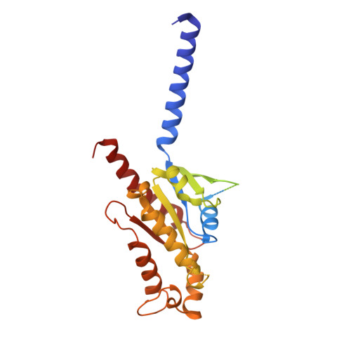
Guanine nucleotide-binding protein G(i) subunit alpha-1354Homo sapiensMutation(s): 0 
Gene Names: GNAI1
EC: 3.6.5
Membrane Entity: Yes 
UniProt & NIH Common Fund Data Resources
Find proteins for P63096 (Homo sapiens)
Explore P63096 
Go to UniProtKB:  P63096
PHAROS:  P63096
GTEx:  ENSG00000127955 
Entity Groups  
Sequence Clusters30% Identity50% Identity70% Identity90% Identity95% Identity100% Identity
UniProt GroupP63096
Sequence Annotations
Expand
  • Reference Sequence
Find similar proteins by:  (by identity cutoff)  |  3D Structure
Entity ID: 2
MoleculeChains Sequence LengthOrganismDetailsImage
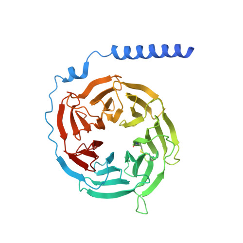
Guanine nucleotide-binding protein G(I)/G(S)/G(T) subunit beta-1358Homo sapiensMutation(s): 0 
Gene Names: GNB1
UniProt & NIH Common Fund Data Resources
Find proteins for P62873 (Homo sapiens)
Explore P62873 
Go to UniProtKB:  P62873
PHAROS:  P62873
GTEx:  ENSG00000078369 
Entity Groups  
Sequence Clusters30% Identity50% Identity70% Identity90% Identity95% Identity100% Identity
UniProt GroupP62873
Sequence Annotations
Expand
  • Reference Sequence
Find similar proteins by:  (by identity cutoff)  |  3D Structure
Entity ID: 3
MoleculeChains Sequence LengthOrganismDetailsImage
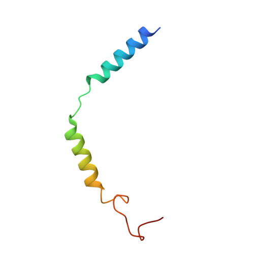
Guanine nucleotide-binding protein G(I)/G(S)/G(O) subunit gamma-258Homo sapiensMutation(s): 0 
Gene Names: GNG2
Membrane Entity: Yes 
UniProt & NIH Common Fund Data Resources
Find proteins for P59768 (Homo sapiens)
Explore P59768 
Go to UniProtKB:  P59768
PHAROS:  P59768
GTEx:  ENSG00000186469 
Entity Groups  
Sequence Clusters30% Identity50% Identity70% Identity90% Identity95% Identity100% Identity
UniProt GroupP59768
Sequence Annotations
Expand
  • Reference Sequence
Find similar proteins by:  (by identity cutoff)  |  3D Structure
Entity ID: 4
MoleculeChains Sequence LengthOrganismDetailsImage
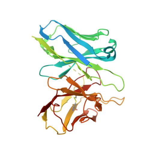
scFvD [auth S]285Homo sapiensMutation(s): 0 
Entity Groups  
Sequence Clusters30% Identity50% Identity70% Identity90% Identity95% Identity100% Identity
Sequence Annotations
Expand
  • Reference Sequence
Find similar proteins by:  (by identity cutoff)  |  3D Structure
Entity ID: 5
MoleculeChains Sequence LengthOrganismDetailsImage
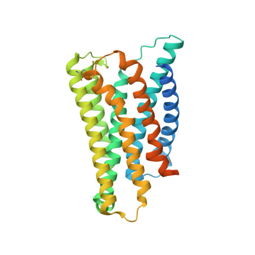
Mas-related G-protein coupled receptor member X2E [auth R]330Homo sapiensMutation(s): 0 
Gene Names: MRGPRX2MRGX2
Membrane Entity: Yes 
UniProt & NIH Common Fund Data Resources
Find proteins for Q96LB1 (Homo sapiens)
Explore Q96LB1 
Go to UniProtKB:  Q96LB1
PHAROS:  Q96LB1
GTEx:  ENSG00000183695 
Entity Groups  
Sequence Clusters30% Identity50% Identity70% Identity90% Identity95% Identity100% Identity
UniProt GroupQ96LB1
Sequence Annotations
Expand
  • Reference Sequence

Find similar proteins by:  Sequence   |   3D Structure  

Entity ID: 6
MoleculeChains Sequence LengthOrganismDetailsImage
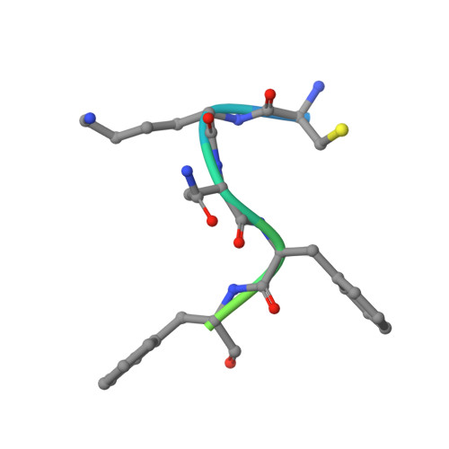
circular cortistatin-14F [auth L]14Homo sapiensMutation(s): 0 
Gene Names: CORTUNQ307/PRO350
Membrane Entity: Yes 
UniProt & NIH Common Fund Data Resources
Find proteins for O00230 (Homo sapiens)
Explore O00230 
Go to UniProtKB:  O00230
PHAROS:  O00230
GTEx:  ENSG00000241563 
Entity Groups  
Sequence Clusters30% Identity50% Identity70% Identity90% Identity95% Identity100% Identity
UniProt GroupO00230
Sequence Annotations
Expand
  • Reference Sequence
Small Molecules
Ligands 1 Unique
IDChains Name / Formula / InChI Key2D Diagram3D Interactions
CLR
Query on CLR

Download Ideal Coordinates CCD File 
G [auth R]CHOLESTEROL
C27 H46 O
HVYWMOMLDIMFJA-DPAQBDIFSA-N
Experimental Data & Validation

Experimental Data

  • Method: ELECTRON MICROSCOPY
  • Resolution: 3.22 Å
  • Aggregation State: PARTICLE 
  • Reconstruction Method: SINGLE PARTICLE 

Structure Validation

View Full Validation Report


Entry History & Funding Information

Deposition Data


Funding OrganizationLocationGrant Number
National Natural Science Foundation of China (NSFC)China81773704, 92057121, 31900936, 31971195

Revision History  (Full details and data files)

  • Version 1.0: 2021-12-01
    Type: Initial release
  • Version 1.1: 2022-07-20
    Changes: Database references, Structure summary
  • Version 1.2: 2024-10-30
    Changes: Data collection, Structure summary