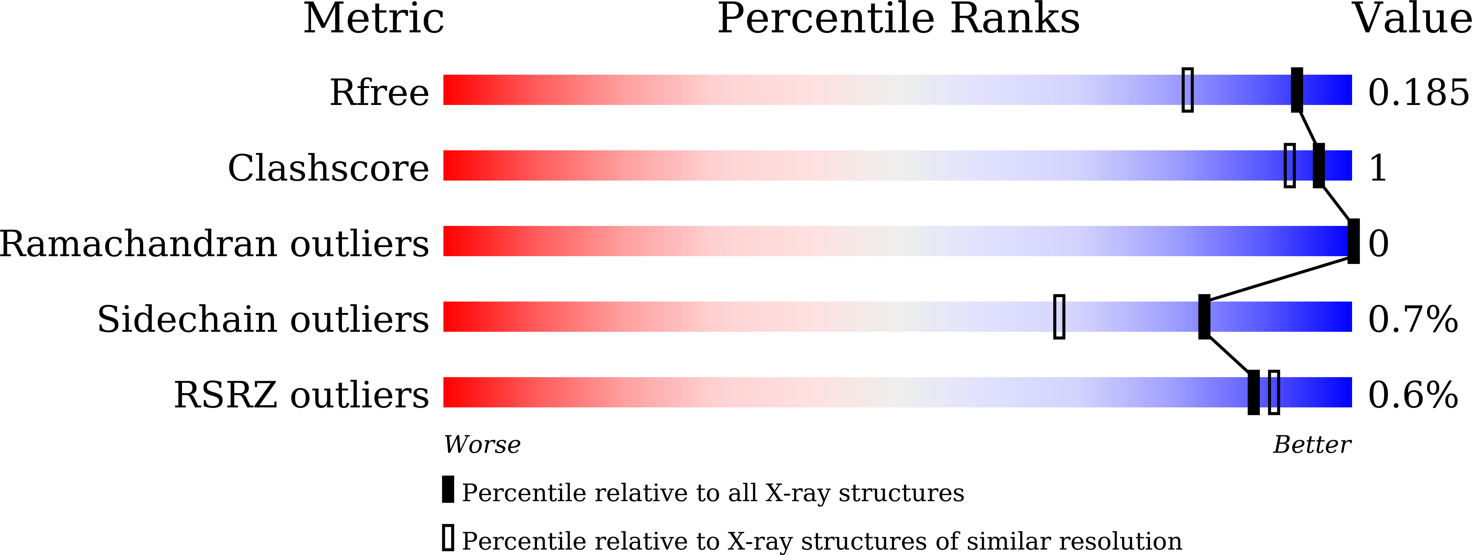
7VB3

Crystal structure of hydroxynitrile lyase from Linum usitatissimum


Experimental Data Snapshot

  • Method: X-RAY DIFFRACTION
  • Resolution: 1.48 Å
  • R-Value Free: 0.183 
  • R-Value Work: 0.156 
  • R-Value Observed: 0.157 

Starting Model: experimental
View more details

wwPDB Validation   3D Report Full Report


Ligand Structure Quality Assessment 


This is version 1.3 of the entry. See complete history


Literature

Structural characterization of Linum usitatissimum hydroxynitrile lyase: A new cyanohydrin decomposition mechanism involving a cyano-zinc complex.

Zheng, D.Nakabayashi, M.Asano, Y.

(2022) J Biol Chem 298: 101650-101650

  • DOI: https://doi.org/10.1016/j.jbc.2022.101650
  • Primary Citation of Related Structures:  
    7VB3, 7VB5, 7VB6

  • PubMed Abstract: 

    Hydroxynitrile lyase from Linum usitatissimum (LuHNL) is an enzyme involved in the catabolism of cyanogenic glycosides to release hydrogen cyanide upon tissue damage. This enzyme strictly conserves the substrate- and NAD(H)-binding domains of Zn 2+ -containing alcohol dehydrogenase (ADH); however, there is no evidence suggesting that LuHNL possesses ADH activity. Herein, we determined the ligand-free 3D structure of LuHNL and its complex with acetone cyanohydrin and (R)-2-butanone cyanohydrin using X-ray crystallography. These structures reveal that an A-form NAD + is tightly but not covalently bound to each subunit of LuHNL. The restricted movement of the NAD+ molecule is due to the "sandwich structure" on the adenine moiety of NAD + . Moreover, the structures and mutagenesis analysis reveal a novel reaction mechanism for cyanohydrin decomposition involving the cyano-zinc complex and hydrogen-bonded interaction of the hydroxyl group of cyanohydrin with Glu323/Thr65 and H 2 O/Lys162 of LuHNL. The deprotonated Lys162 and protonated Glu323 residues are presumably stabilized by a partially desolvated microenvironment. In summary, the substrate binding geometry of LuHNL provides insights into the differences in activities of LuHNL and ADH, and identifying this novel reaction mechanism is an important contribution to the study of hydroxynitrile lyases.


  • Organizational Affiliation

    Biotechnology Research Center and Department of Biotechnology, Toyama Prefectural University, Imizu, Toyama, Japan.


Macromolecules
Find similar proteins by:  (by identity cutoff)  |  3D Structure
Entity ID: 1
MoleculeChains Sequence LengthOrganismDetailsImage
Aliphatic (R)-hydroxynitrile lyase
A, B, C, D
443Linum usitatissimumMutation(s): 0 
EC: 4.1.2.46
UniProt
Find proteins for P93243 (Linum usitatissimum)
Explore P93243 
Go to UniProtKB:  P93243
Entity Groups  
Sequence Clusters30% Identity50% Identity70% Identity90% Identity95% Identity100% Identity
UniProt GroupP93243
Sequence Annotations
Expand
  • Reference Sequence
Small Molecules
Ligands 6 Unique
IDChains Name / Formula / InChI Key2D Diagram3D Interactions
NAD (Subject of Investigation/LOI)
Query on NAD

Download Ideal Coordinates CCD File 
E [auth A],
EA [auth C],
KA [auth D],
R [auth B]
NICOTINAMIDE-ADENINE-DINUCLEOTIDE
C21 H27 N7 O14 P2
BAWFJGJZGIEFAR-NNYOXOHSSA-N
BTB
Query on BTB

Download Ideal Coordinates CCD File 
DA [auth B],
P [auth A],
Q [auth A]
2-[BIS-(2-HYDROXY-ETHYL)-AMINO]-2-HYDROXYMETHYL-PROPANE-1,3-DIOL
C8 H19 N O5
OWMVSZAMULFTJU-UHFFFAOYSA-N
GOL (Subject of Investigation/LOI)
Query on GOL

Download Ideal Coordinates CCD File 
H [auth A]
HA [auth C]
I [auth A]
J [auth A]
K [auth A]
H [auth A],
HA [auth C],
I [auth A],
J [auth A],
K [auth A],
NA [auth D],
OA [auth D],
U [auth B],
V [auth B],
W [auth B],
X [auth B]
GLYCEROL
C3 H8 O3
PEDCQBHIVMGVHV-UHFFFAOYSA-N
ZN (Subject of Investigation/LOI)
Query on ZN

Download Ideal Coordinates CCD File 
F [auth A]
FA [auth C]
G [auth A]
GA [auth C]
LA [auth D]
F [auth A],
FA [auth C],
G [auth A],
GA [auth C],
LA [auth D],
MA [auth D],
S [auth B],
T [auth B]
ZINC ION
Zn
PTFCDOFLOPIGGS-UHFFFAOYSA-N
EDO
Query on EDO

Download Ideal Coordinates CCD File 
AA [auth B]
BA [auth B]
CA [auth B]
JA [auth C]
M [auth A]
AA [auth B],
BA [auth B],
CA [auth B],
JA [auth C],
M [auth A],
N [auth A],
O [auth A],
QA [auth D],
Z [auth B]
1,2-ETHANEDIOL
C2 H6 O2
LYCAIKOWRPUZTN-UHFFFAOYSA-N
MG
Query on MG

Download Ideal Coordinates CCD File 
IA [auth C],
L [auth A],
PA [auth D],
Y [auth B]
MAGNESIUM ION
Mg
JLVVSXFLKOJNIY-UHFFFAOYSA-N
Modified Residues  1 Unique
IDChains TypeFormula2D DiagramParent
SNC
Query on SNC
A, B, C, D
L-PEPTIDE LINKINGC3 H6 N2 O3 SCYS
Experimental Data & Validation

Experimental Data

  • Method: X-RAY DIFFRACTION
  • Resolution: 1.48 Å
  • R-Value Free: 0.183 
  • R-Value Work: 0.156 
  • R-Value Observed: 0.157 
  • Space Group: P 1 21 1
Unit Cell:
Length ( Å )Angle ( ˚ )
a = 94.12α = 90
b = 52.18β = 95.01
c = 168.51γ = 90
Software Package:
Software NamePurpose
REFMACrefinement
SCALAdata scaling
PDB_EXTRACTdata extraction
Cootmodel building
MOSFLMdata reduction

Structure Validation

View Full Validation Report


Ligand Structure Quality Assessment 


Entry History & Funding Information

Deposition Data


Funding OrganizationLocationGrant Number
Japan Society for the Promotion of Science (JSPS)Japan--

Revision History  (Full details and data files)

  • Version 1.0: 2022-02-09
    Type: Initial release
  • Version 1.1: 2022-03-16
    Changes: Database references
  • Version 1.2: 2023-11-29
    Changes: Data collection, Refinement description
  • Version 1.3: 2024-10-16
    Changes: Structure summary