7V9E | pdb_00007v9e

Crystal structure of a methyl transferase ribozyme

  • Classification: RNA
  • Organism(s): Homo sapiens
  • Mutation(s): No 

  • Deposited: 2021-08-25 Released: 2022-03-23 
  • Deposition Author(s): Deng, J., Lilley, D.M.J., Huang, L.
  • Funding Organization(s): Ministry of Education (MoE, China), Cancer Research UK

Experimental Data Snapshot

  • Method: X-RAY DIFFRACTION
  • Resolution: 2.30 Å
  • R-Value Free: 
    0.271 (Depositor), 0.283 (DCC) 
  • R-Value Work: 
    0.244 (Depositor), 0.252 (DCC) 
  • R-Value Observed: 
    0.245 (Depositor) 

wwPDB Validation 3D Report Full Report

Validation slider image for 7V9E

Ligand Structure Quality Assessment 


This is version 1.3 of the entry. See complete history

Literature

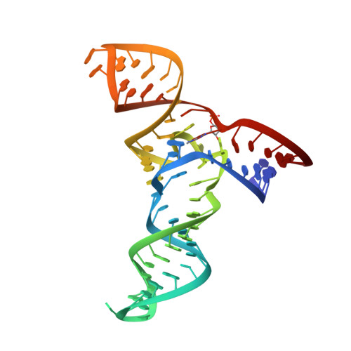
Structure and mechanism of a methyltransferase ribozyme.

Deng, J.Wilson, T.J.Wang, J.Peng, X.Li, M.Lin, X.Liao, W.Lilley, D.M.J.Huang, L.

(2022) Nat Chem Biol 18: 556-564

  • DOI: https://doi.org/10.1038/s41589-022-00982-z
  • Primary Citation Related Structures: 
    7V9E

  • PubMed Abstract: 

    Known ribozymes in contemporary biology perform a limited range of chemical catalysis, but in vitro selection has generated species that catalyze a broader range of chemistry; yet, there have been few structural and mechanistic studies of selected ribozymes. A ribozyme has recently been selected that can catalyze a site-specific methyl transfer reaction. We have solved the crystal structure of this ribozyme at a resolution of 2.3 Å, showing how the RNA folds to generate a very specific binding site for the methyl donor substrate. The structure immediately suggests a catalytic mechanism involving a combination of proximity and orientation and nucleobase-mediated general acid catalysis. The mechanism is supported by the pH dependence of the rate of catalysis. A selected methyltransferase ribozyme can thus use a relatively sophisticated catalytic mechanism, broadening the range of known RNA-catalyzed chemistry.


  • Organizational Affiliation
    • Guangdong Provincial Key Laboratory of Malignant Tumor Epigenetics and Gene Regulation, Guangdong-Hong Kong Joint Laboratory for RNA Medicine, Sun Yat-Sen Memorial Hospital, Sun Yat-Sen University, Guangzhou, China.

Macromolecule Content 

  • Total Structure Weight: 23.32 kDa 
  • Atom Count: 1,494 
  • Modeled Residue Count: 68 
  • Deposited Residue Count: 68 
  • Unique nucleic acid chains: 1

Macromolecules

Find similar nucleic acids by:  (by identity cutoff) 
Entity ID: 1
MoleculeChains LengthOrganismImage
RNA (68-MER)68Homo sapiens
Sequence Annotations
Expand
Reference Sequence

Small Molecules

Ligands 3 Unique
IDChains Name / Formula / InChI Key2D Diagram3D Interactions
GUN
(Subject of Investigation/LOI)

Query on GUN


Download:Ideal Coordinates CCD File
B [auth A]GUANINE
C5 H5 N5 O
UYTPUPDQBNUYGX-UHFFFAOYSA-N
BA

Query on BA


Download:Ideal Coordinates CCD File
C [auth A]
D [auth A]
E [auth A]
F [auth A]
G [auth A]
C [auth A],
D [auth A],
E [auth A],
F [auth A],
G [auth A],
H [auth A],
I [auth A],
J [auth A],
K [auth A]
BARIUM ION
Ba
XDFCIPNJCBUZJN-UHFFFAOYSA-N
NA

Query on NA


Download:Ideal Coordinates CCD File
L [auth A],
M [auth A]
SODIUM ION
Na
FKNQFGJONOIPTF-UHFFFAOYSA-N

Experimental Data & Validation

Experimental Data

  • Method: X-RAY DIFFRACTION
  • Resolution: 2.30 Å
  • R-Value Free:  0.271 (Depositor), 0.283 (DCC) 
  • R-Value Work:  0.244 (Depositor), 0.252 (DCC) 
  • R-Value Observed: 0.245 (Depositor) 
Space Group: P 2 2 21
Unit Cell:
Length ( Å )Angle ( ˚ )
a = 50.381α = 90
b = 52.507β = 90
c = 99.939γ = 90
Software Package:
Software NamePurpose
PHENIXrefinement
ADDREFdata reduction
Aimlessdata scaling
PHENIXphasing

Structure Validation

View Full Validation Report


Ligand Structure Quality Assessment 


Entry History 

& Funding Information

Deposition Data


Funding OrganizationLocationGrant Number
Ministry of Education (MoE, China)United Kingdom--
Cancer Research UKUnited KingdomA18604

Revision History  (Full details and data files)

  • Version 1.0: 2022-03-23
    Type: Initial release
  • Version 1.1: 2022-03-30
    Changes: Database references
  • Version 1.2: 2022-05-11
    Changes: Database references
  • Version 1.3: 2024-05-29
    Changes: Data collection