


The structural basis for 2'-5'/3'-5'-cGAMP synthesis by cGAS
Wu, S., Gabelli, S.B., Sohn, J.(2024) Nat Commun 15: 4012
Experimental Data Snapshot
Starting Model: experimental
View more details
(2024) Nat Commun 15: 4012
Entity ID: 1 | |||||
|---|---|---|---|---|---|
| Molecule | Chains | Sequence Length | Organism | Details | Image |
| Cyclic GMP-AMP synthase | A, B [auth C] | 364 | Mus musculus | Mutation(s): 2 Gene Names: Cgas, Mb21d1 EC: 2.7.7.86 | 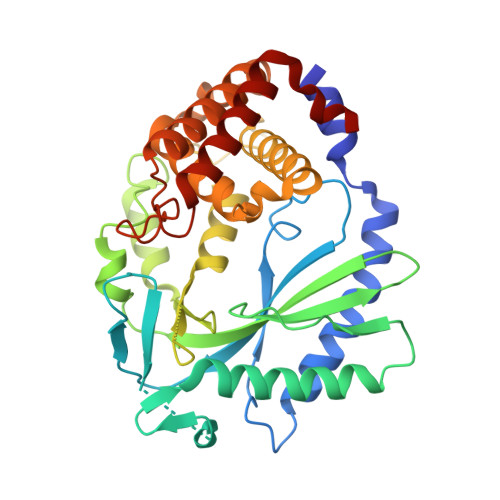 |
UniProt & NIH Common Fund Data Resources | |||||
IMPC: MGI:2442261 | |||||
Entity Groups | |||||
| Sequence Clusters | 30% Identity50% Identity70% Identity90% Identity95% Identity100% Identity | ||||
| UniProt Group | Q8C6L5 | ||||
Sequence AnnotationsExpand | |||||
Reference Sequence | |||||
Entity ID: 2 | ||||
| Molecule | Chains | Length | Organism | Image |
|---|---|---|---|---|
| Palindromic DNA18 | C [auth E], D [auth F], E [auth I], F [auth J] | 18 | synthetic construct | 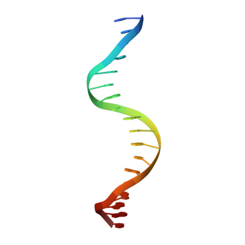 |
Sequence AnnotationsExpand | ||||
Reference Sequence | ||||
| Ligands 4 Unique | |||||
|---|---|---|---|---|---|
| ID | Chains | Name / Formula / InChI Key | 2D Diagram | 3D Interactions | |
| GTP (Subject of Investigation/LOI) Download:Ideal Coordinates CCD File | J [auth A], N [auth C] | GUANOSINE-5'-TRIPHOSPHATE C10 H16 N5 O14 P3 XKMLYUALXHKNFT-UUOKFMHZSA-N |  | ||
| AMP (Subject of Investigation/LOI) Download:Ideal Coordinates CCD File | I [auth A], M [auth C] | ADENOSINE MONOPHOSPHATE C10 H14 N5 O7 P UDMBCSSLTHHNCD-KQYNXXCUSA-N |  | ||
| ZN Download:Ideal Coordinates CCD File | G [auth A], K [auth C] | ZINC ION Zn PTFCDOFLOPIGGS-UHFFFAOYSA-N |  | ||
| MN (Subject of Investigation/LOI) Download:Ideal Coordinates CCD File | H [auth A], L [auth C] | MANGANESE (II) ION Mn WAEMQWOKJMHJLA-UHFFFAOYSA-N |  | ||
| Length ( Å ) | Angle ( ˚ ) |
|---|---|
| a = 78.475 | α = 90 |
| b = 99.474 | β = 90 |
| c = 142.088 | γ = 90 |
| Software Name | Purpose |
|---|---|
| XDS | data reduction |
| Aimless | data scaling |
| MOLREP | phasing |
| REFMAC | refinement |
| PDB-REDO | refinement |
| PDB_EXTRACT | data extraction |
| Funding Organization | Location | Grant Number |
|---|---|---|
| National Institutes of Health/National Institute of General Medical Sciences (NIH/NIGMS) | United States | -- |


