7USN | pdb_00007usn

Crystal structure of ferritin 1 from Caenorhabditis elegans, FTN-1


Experimental Data Snapshot

  • Method: X-RAY DIFFRACTION
  • Resolution: 1.79 Å
  • R-Value Free: 
    0.189 (Depositor), 0.196 (DCC) 
  • R-Value Work: 
    0.166 (Depositor), 0.174 (DCC) 

wwPDB Validation 3D Report Full Report

Validation slider image for 7USN

This is version 1.2 of the entry. See complete history

Literature

Biochemical Characterization of Caenorhabditis elegans Ferritins.

Mubarak, S.S.M.Malcolm, T.R.Brown, H.G.Hanssen, E.Maher, M.J.McColl, G.Jameson, G.N.L.

(2023) Biochemistry 62: 1484-1496

  • DOI: https://doi.org/10.1021/acs.biochem.3c00005
  • Primary Citation Related Structures: 
    7URH, 7USN

  • PubMed Abstract: 

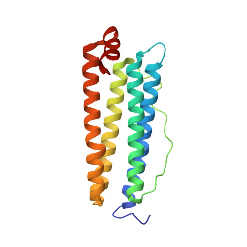
    The nematode Caenorhabditis elegans contains genes for two types of ferritin ( ftn-1 and ftn-2 ) that express FTN-1 and FTN-2. We have expressed and purified both proteins and characterized them by X-ray crystallography, cryo-electron microscopy, transmission electron microscopy, dynamic light scattering, and kinetically by oxygen electrode and UV-vis spectroscopy. Both show ferroxidase activity, but although they have identical ferroxidase active sites, FTN-2 is shown to react approximately 10 times faster than FTN-1, with L-type ferritin character over longer time periods. We hypothesize that the large variation in rate may be due to differences in the three- and four-fold channels into the interior of the protein 24-mer. FTN-2 is shown to have a wider entrance into the three-fold channel than FTN-1. Additionally, the charge gradient through the channel of FTN-2 is more pronounced, with Asn and Gln residues in FTN-1 replaced by Asp and Glu residues in FTN-2. Both FTN-1 and FTN-2 have an Asn residue near the ferroxidase active site that is a Val in most other species, including human H ferritin. This Asn residue has been observed before in ferritin from the marine pennate diatom Pseudo-mitzchia multiseries. By replacing this Asn residue with a Val in FTN-2, we show that the reactivity decreases over long time scales. We therefore propose that Asn106 is involved in iron transport from the ferroxidase active site to the central cavity of the protein.


  • Organizational Affiliation
    • School of Chemistry, Bio21 Molecular Science and Biotechnology Institute, The University of Melbourne, 30 Flemington Road, Parkville, Victoria 3010, Australia.

Macromolecule Content 

  • Total Structure Weight: 313.83 kDa 
  • Atom Count: 24,606 
  • Modeled Residue Count: 2,688 
  • Deposited Residue Count: 2,688 
  • Unique protein chains: 1

Macromolecules

Find similar proteins by:|  3D Structure
Entity ID: 1
MoleculeChains  Sequence LengthOrganismDetailsImage
Ferritin168Caenorhabditis elegansMutation(s): 0 
Gene Names: ftn-1C54F6.14CELE_C54F6.14
EC: 1.16.3.1
UniProt
Find proteins for O16453 (Caenorhabditis elegans)
Explore O16453 
Go to UniProtKB:  O16453
Entity Groups
Sequence Clusters30% Identity50% Identity70% Identity90% Identity95% Identity100% Identity
UniProt GroupO16453
Sequence Annotations
Expand
Reference Sequence

Small Molecules

Ligands 3 Unique
IDChains Name / Formula / InChI Key2D Diagram3D Interactions
SO4

Query on SO4


Download:Ideal Coordinates CCD File
CB [auth J]
FB [auth K]
GA [auth D]
JA [auth E]
JB [auth L]
CB [auth J],
FB [auth K],
GA [auth D],
JA [auth E],
JB [auth L],
NA [auth F],
RA [auth G],
RB [auth P],
T [auth A],
U [auth A],
UA [auth O],
V [auth A],
WA [auth H],
YA [auth I],
Z [auth B]
SULFATE ION
O4 S
QAOWNCQODCNURD-UHFFFAOYSA-L
GOL

Query on GOL


Download:Ideal Coordinates CCD File
AB [auth J]
BA [auth C]
BB [auth J]
CA [auth C]
EA [auth D]
AB [auth J],
BA [auth C],
BB [auth J],
CA [auth C],
EA [auth D],
EB [auth K],
FA [auth D],
HB [auth L],
IA [auth E],
IB [auth L],
LA [auth F],
MA [auth F],
MB [auth N],
NB [auth N],
PA [auth G],
PB [auth P],
Q [auth A],
QA [auth G],
QB [auth P],
R [auth A],
S [auth A],
TA [auth O],
X [auth B],
Y [auth B]
GLYCEROL
C3 H8 O3
PEDCQBHIVMGVHV-UHFFFAOYSA-N
FE

Query on FE


Download:Ideal Coordinates CCD File
AA [auth B]
DA [auth C]
DB [auth J]
GB [auth K]
HA [auth D]
AA [auth B],
DA [auth C],
DB [auth J],
GB [auth K],
HA [auth D],
KA [auth E],
KB [auth L],
LB [auth M],
OA [auth F],
OB [auth N],
SA [auth G],
SB [auth P],
VA [auth O],
W [auth A],
XA [auth H],
ZA [auth I]
FE (III) ION
Fe
VTLYFUHAOXGGBS-UHFFFAOYSA-N

Experimental Data & Validation

Experimental Data

  • Method: X-RAY DIFFRACTION
  • Resolution: 1.79 Å
  • R-Value Free:  0.189 (Depositor), 0.196 (DCC) 
  • R-Value Work:  0.166 (Depositor), 0.174 (DCC) 
Space Group: P 2 3
Unit Cell:
Length ( Å )Angle ( ˚ )
a = 227.763α = 90
b = 227.763β = 90
c = 227.763γ = 90
Software Package:
Software NamePurpose
REFMACrefinement
XDSdata reduction
Aimlessdata scaling
SHELXDphasing

Structure Validation

View Full Validation Report


Entry History 

& Funding Information

Deposition Data


Funding OrganizationLocationGrant Number
Australian Research Council (ARC)Australia--

Revision History  (Full details and data files)

  • Version 1.0: 2023-04-26
    Type: Initial release
  • Version 1.1: 2023-05-17
    Changes: Database references
  • Version 1.2: 2024-05-22
    Changes: Data collection