


Evolution of Antibody Reactivity against EBV EBNA1 to Molecular Mimicry with GlialCAM
Lanz, T.V., Robinson, W.H., Fernandez, D.To be published.
Experimental Data Snapshot
Starting Model: experimental
View more details
Entity ID: 1 | |||||
|---|---|---|---|---|---|
| Molecule | Chains | Sequence Length | Organism | Details | Image |
| Fab MS39p2w174 Light Chain | A, D [auth G], F [auth I], H [auth D] | 219 | Homo sapiens | Mutation(s): 0 | 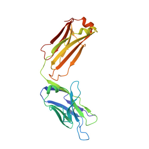 |
Entity ID: 2 | |||||
|---|---|---|---|---|---|
| Molecule | Chains | Sequence Length | Organism | Details | Image |
| Fab MS39p2w174 Heavy Chain | B, E [auth H], G [auth J], I [auth E] | 219 | Homo sapiens | Mutation(s): 0 | 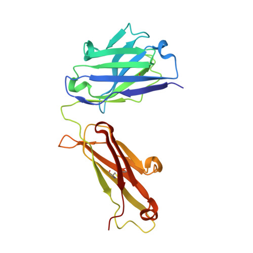 |
Entity ID: 3 | |||||
|---|---|---|---|---|---|
| Molecule | Chains | Sequence Length | Organism | Details | Image |
| Hepatocyte cell adhesion molecule | C, J [auth F] | 20 | Homo sapiens | Mutation(s): 0 Gene Names: HEPACAM | 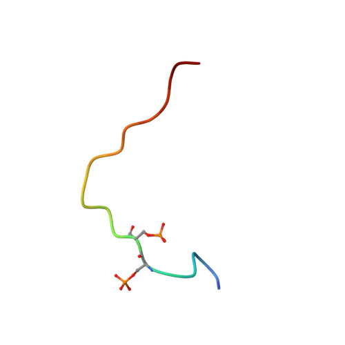 |
UniProt & NIH Common Fund Data Resources | |||||
PHAROS: Q14CZ8 GTEx: ENSG00000165478 | |||||
Entity Groups | |||||
| Sequence Clusters | 30% Identity50% Identity70% Identity90% Identity95% Identity100% Identity | ||||
| UniProt Group | Q14CZ8 | ||||
Sequence AnnotationsExpand | |||||
Reference Sequence | |||||
| Ligands 2 Unique | |||||
|---|---|---|---|---|---|
| ID | Chains | Name / Formula / InChI Key | 2D Diagram | 3D Interactions | |
| SO4 (Subject of Investigation/LOI) Download:Ideal Coordinates CCD File | K [auth A], Q [auth I] | SULFATE ION O4 S QAOWNCQODCNURD-UHFFFAOYSA-L |  | ||
| CL (Subject of Investigation/LOI) Download:Ideal Coordinates CCD File | L [auth B] M [auth G] N [auth G] O [auth H] P [auth H] | CHLORIDE ION Cl VEXZGXHMUGYJMC-UHFFFAOYSA-M |  | ||
| Modified Residues 1 Unique | |||||
|---|---|---|---|---|---|
| ID | Chains | Type | Formula | 2D Diagram | Parent |
| SEP Query on SEP | C, J [auth F] | L-PEPTIDE LINKING | C3 H8 N O6 P |  | SER |
| Length ( Å ) | Angle ( ˚ ) |
|---|---|
| a = 195.027 | α = 90 |
| b = 99.514 | β = 112.811 |
| c = 136.414 | γ = 90 |
| Software Name | Purpose |
|---|---|
| REFMAC | refinement |
| PHENIX | refinement |
| XDS | data reduction |
| SCALA | data scaling |
| PHASER | phasing |
| Funding Organization | Location | Grant Number |
|---|---|---|
| National Institutes of Health/National Institute Of Allergy and Infectious Diseases (NIH/NIAID) | United States | AR063676 |


