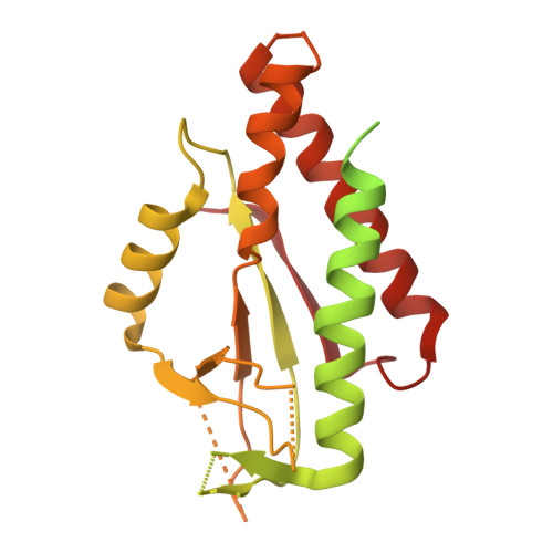
7UNA

SfSTING with cGAMP (masked)


Experimental Data Snapshot

  • Method: ELECTRON MICROSCOPY
  • Resolution: 4.00 Å
  • Aggregation State: FILAMENT 
  • Reconstruction Method: SINGLE PARTICLE 

wwPDB Validation   3D Report Full Report


This is version 1.3 of the entry. See complete history


Literature

Cryo-EM structure of an active bacterial TIR-STING filament complex.

Morehouse, B.R.Yip, M.C.J.Keszei, A.F.A.McNamara-Bordewick, N.K.Shao, S.Kranzusch, P.J.

(2022) Nature 608: 803-807

  • DOI: https://doi.org/10.1038/s41586-022-04999-1
  • Primary Citation of Related Structures:  
    7UN8, 7UN9, 7UNA

  • PubMed Abstract: 

    Stimulator of interferon genes (STING) is an antiviral signalling protein that is broadly conserved in both innate immunity in animals and phage defence in prokaryotes 1-4 . Activation of STING requires its assembly into an oligomeric filament structure through binding of a cyclic dinucleotide 4-13 , but the molecular basis of STING filament assembly and extension remains unknown. Here we use cryogenic electron microscopy to determine the structure of the active Toll/interleukin-1 receptor (TIR)-STING filament complex from a Sphingobacterium faecium cyclic-oligonucleotide-based antiphage signalling system (CBASS) defence operon. Bacterial TIR-STING filament formation is driven by STING interfaces that become exposed on high-affinity recognition of the cognate cyclic dinucleotide signal c-di-GMP. Repeating dimeric STING units stack laterally head-to-head through surface interfaces, which are also essential for human STING tetramer formation and downstream immune signalling in mammals 5 . The active bacterial TIR-STING structure reveals further cross-filament contacts that brace the assembly and coordinate packing of the associated TIR NADase effector domains at the base of the filament to drive NAD + hydrolysis. STING interface and cross-filament contacts are essential for cell growth arrest in vivo and reveal a stepwise mechanism of activation whereby STING filament assembly is required for subsequent effector activation. Our results define the structural basis of STING filament formation in prokaryotic antiviral signalling.


  • Organizational Affiliation

    Department of Microbiology, Harvard Medical School, Boston, MA, USA.


Macromolecules
Find similar proteins by:  (by identity cutoff)  |  3D Structure
Entity ID: 1
MoleculeChains Sequence LengthOrganismDetailsImage
CD-NTase-associated protein 12
A, B, C, D, E
A, B, C, D, E, F, G, H
331Sphingobacterium faeciumMutation(s): 0 
Gene Names: cap12C8N37_104320SF1_08920
EC: 3.2.2.5
UniProt
Find proteins for A0A2T5Y4G4 (Sphingobacterium faecium (strain DSM 11690 / JCM 21820 / NBRC 15299 / NCIMB 13408 / KS 0470))
Explore A0A2T5Y4G4 
Go to UniProtKB:  A0A2T5Y4G4
Entity Groups  
Sequence Clusters30% Identity50% Identity70% Identity90% Identity95% Identity100% Identity
UniProt GroupA0A2T5Y4G4
Sequence Annotations
Expand
  • Reference Sequence
Small Molecules
Experimental Data & Validation

Experimental Data

  • Method: ELECTRON MICROSCOPY
  • Resolution: 4.00 Å
  • Aggregation State: FILAMENT 
  • Reconstruction Method: SINGLE PARTICLE 

Structure Validation

View Full Validation Report


Entry History & Funding Information

Deposition Data


Funding OrganizationLocationGrant Number
The Pew Charitable TrustsUnited States--
Burroughs Wellcome FundUnited States--
The G. Harold and Leila Y. Mathers FoundationUnited States--
The Mark FoundationUnited States--
Parker Institute for Cancer ImmunotherapyUnited States--
National Institutes of Health/National Institute of General Medical Sciences (NIH/NIGMS)United States1DP2GM146250
The Vallee FoundationUnited States--
David and Lucile Packard FoundationUnited States--
National Institutes of Health/National Institute of General Medical Sciences (NIH/NIGMS)United States1DP2GM137415
National Institutes of Health/National Institute of General Medical Sciences (NIH/NIGMS)United StatesF32GM133063
American Heart AssociationUnited States287375208

Revision History  (Full details and data files)

  • Version 1.0: 2022-07-27
    Type: Initial release
  • Version 1.1: 2022-08-03
    Changes: Database references
  • Version 1.2: 2022-09-07
    Changes: Database references
  • Version 1.3: 2024-02-14
    Changes: Data collection