7ULT | pdb_00007ult

Crystal Structure of SARS-CoV-2 Nsp16/10 Heterodimer Apo-Form.


Experimental Data Snapshot

  • Method: X-RAY DIFFRACTION
  • Resolution: 1.90 Å
  • R-Value Free: 
    0.188 (Depositor), 0.194 (DCC) 
  • R-Value Work: 
    0.164 (Depositor), 0.171 (DCC) 
  • R-Value Observed: 
    0.166 (Depositor) 

Starting Model: experimental
View more details

wwPDB Validation 3D Report Full Report

Validation slider image for 7ULT

This is version 1.2 of the entry. See complete history

Literature

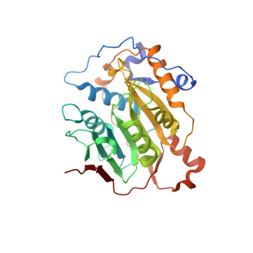
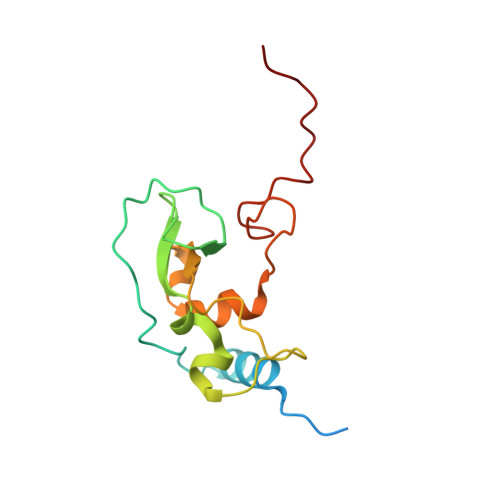
S -adenosylhomocysteine analogs selectively suppress pan-coronavirus replication by inhibition of nsp14 methyltransferase.

Rosas-Lemus, M.Athe, S.Minasov, G.Pattie, J.A.Brunzelle, J.S.Chau, I.Li, F.Vedadi, M.Ma, H.Ramanathan, A.Becker, M.E.Hope, T.J.Abdelkarim, H.Grudzien, P.Gaponenko, V.Montgomery, J.E.Moellering, R.E.Rawal, V.H.Satchell, K.J.F.

(2025) ACS Med Chem Lett 

  • DOI: https://doi.org/10.1021/acsmedchemlett.5c00510
  • Primary Citation Related Structures: 
    7ULT

  • PubMed Abstract: 

    To address the ongoing threat of SARS-CoV-2 and potential emergence of novel coronaviruses, we employed a comprehensive strategy to identify and synthesize inhibitors of coronavirus methyltransferases with chemical analogs of S -adenosylhomocysteine. Two analogs, designated 4h and 4p , inhibit both mouse hepatitis virus and SARS-CoV-2 replication. Compound 4p was most potent with half-maximal inhibition of biochemical activity at 0.2 μM and antiviral activity at ~20 μM. This compound also has low cytotoxicity and preferentially inhibits nsp14 over nsp16 and human methyltransferases. Furthermore, molecular docking based on a newly determined crystal structure of the apo nsp16-nsp10 complex predicts 4p occupies both the S -adenosylmethione and Gppp binding pockets of nsp14 and nsp16. Selectivity of 4p for nsp14 is likely due to enhanced structural stability of the nsp14 binding pocket relative to nsp16. These findings highlight SAH analogs as scaffolds for pan-coronavirus therapeutics and underscore the value of structure-guided design in antiviral drug discovery.


  • Organizational Affiliation
    • Department of Microbiology-Immunology, Northwestern University Feinberg School of Medicine, Chicago, IL 60611 USA.

Macromolecule Content 

  • Total Structure Weight: 98.35 kDa 
  • Atom Count: 7,433 
  • Modeled Residue Count: 827 
  • Deposited Residue Count: 882 
  • Unique protein chains: 2

Macromolecules

Find similar proteins by:|  3D Structure
Entity ID: 1
MoleculeChains  Sequence LengthOrganismDetailsImage
2'-O-methyltransferase
A, C
300Severe acute respiratory syndrome coronavirus 2Mutation(s): 0 
Gene Names: rep1a-1b
EC: 2.1.1
UniProt
Find proteins for P0DTD1 (Severe acute respiratory syndrome coronavirus 2)
Explore P0DTD1 
Go to UniProtKB:  P0DTD1
Entity Groups
Sequence Clusters30% Identity50% Identity70% Identity90% Identity95% Identity100% Identity
UniProt GroupP0DTD1
Sequence Annotations
Expand
Reference Sequence
Find similar proteins by:|  3D Structure
Entity ID: 2
MoleculeChains  Sequence LengthOrganismDetailsImage
Non-structural protein 10
B, D
141Severe acute respiratory syndrome coronavirus 2Mutation(s): 0 
Gene Names: rep1a-1b
UniProt
Find proteins for P0DTD1 (Severe acute respiratory syndrome coronavirus 2)
Explore P0DTD1 
Go to UniProtKB:  P0DTD1
Entity Groups
Sequence Clusters30% Identity50% Identity70% Identity90% Identity95% Identity100% Identity
UniProt GroupP0DTD1
Sequence Annotations
Expand
Reference Sequence

Small Molecules

Ligands 3 Unique
IDChains Name / Formula / InChI Key2D Diagram3D Interactions
ZN
(Subject of Investigation/LOI)

Query on ZN


Download:Ideal Coordinates CCD File
DA [auth D],
EA [auth D],
N [auth B],
O [auth B]
ZINC ION
Zn
PTFCDOFLOPIGGS-UHFFFAOYSA-N
FMT

Query on FMT


Download:Ideal Coordinates CCD File
AA [auth C]
BA [auth C]
CA [auth C]
G [auth A]
GA [auth D]
AA [auth C],
BA [auth C],
CA [auth C],
G [auth A],
GA [auth D],
H [auth A],
I [auth A],
J [auth A],
K [auth A],
L [auth A],
M [auth A],
U [auth C],
V [auth C],
W [auth C],
X [auth C],
Y [auth C],
Z [auth C]
FORMIC ACID
C H2 O2
BDAGIHXWWSANSR-UHFFFAOYSA-N
NA

Query on NA


Download:Ideal Coordinates CCD File
E [auth A]
F [auth A]
FA [auth D]
P [auth B]
Q [auth B]
E [auth A],
F [auth A],
FA [auth D],
P [auth B],
Q [auth B],
R [auth C],
S [auth C],
T [auth C]
SODIUM ION
Na
FKNQFGJONOIPTF-UHFFFAOYSA-N

Experimental Data & Validation

Experimental Data

  • Method: X-RAY DIFFRACTION
  • Resolution: 1.90 Å
  • R-Value Free:  0.188 (Depositor), 0.194 (DCC) 
  • R-Value Work:  0.164 (Depositor), 0.171 (DCC) 
  • R-Value Observed: 0.166 (Depositor) 
Space Group: P 32 2 1
Unit Cell:
Length ( Å )Angle ( ˚ )
a = 166.681α = 90
b = 166.681β = 90
c = 98.55γ = 120
Software Package:
Software NamePurpose
REFMACrefinement
PDB_EXTRACTdata extraction
HKL-3000data reduction
HKL-3000data scaling
PHASERphasing

Structure Validation

View Full Validation Report


Entry History 

& Funding Information

Deposition Data


Funding OrganizationLocationGrant Number
Not funded--

Revision History  (Full details and data files)

  • Version 1.0: 2022-04-13
    Type: Initial release
  • Version 1.1: 2023-10-18
    Changes: Data collection, Refinement description
  • Version 1.2: 2026-02-11
    Changes: Database references, Structure summary