7UL3 | pdb_00007ul3

CryoEM Structure of Inactive H2R Bound to Famotidine, Nb6M, and NabFab


Experimental Data Snapshot

  • Method: ELECTRON MICROSCOPY
  • Resolution: 3.00 Å
  • Aggregation State: PARTICLE 
  • Reconstruction Method: SINGLE PARTICLE 

wwPDB Validation 3D Report Full Report

Validation slider image for 7UL3

This is version 1.5 of the entry. See complete history

Literature

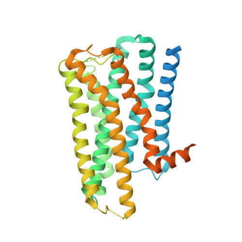
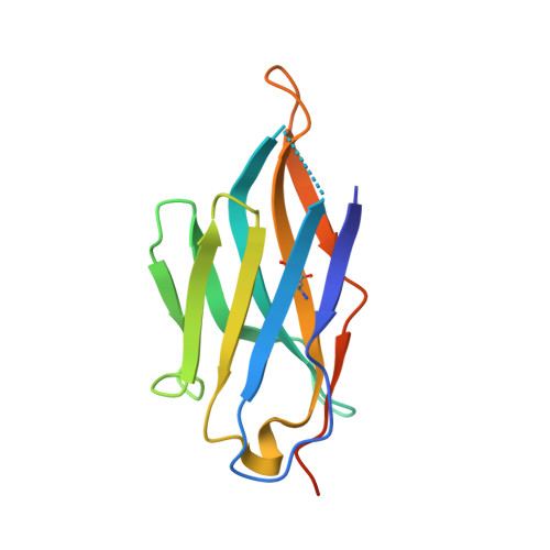
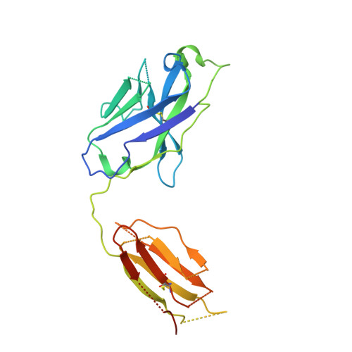
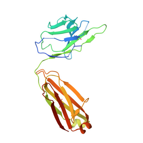
Structure determination of inactive-state GPCRs with a universal nanobody.

Robertson, M.J.Papasergi-Scott, M.M.He, F.Seven, A.B.Meyerowitz, J.G.Panova, O.Peroto, M.C.Che, T.Skiniotis, G.

(2022) Nat Struct Mol Biol 29: 1188-1195

  • DOI: https://doi.org/10.1038/s41594-022-00859-8
  • Primary Citation Related Structures: 
    7UL2, 7UL3, 7UL4, 7UL5

  • PubMed Abstract: 

    Cryogenic electron microscopy (cryo-EM) has widened the field of structure-based drug discovery by allowing for routine determination of membrane protein structures previously intractable. Despite representing one of the largest classes of therapeutic targets, most inactive-state G protein-coupled receptors (GPCRs) have remained inaccessible for cryo-EM because their small size and membrane-embedded nature impedes projection alignment for high-resolution map reconstructions. Here we demonstrate that the same single-chain camelid antibody (nanobody) recognizing a grafted intracellular loop can be used to obtain cryo-EM structures of inactive-state GPCRs at resolutions comparable or better than those obtained by X-ray crystallography. Using this approach, we obtained structures of neurotensin 1 receptor bound to antagonist SR48692, μ-opioid receptor bound to alvimopan, apo somatostatin receptor 2 and histamine receptor 2 bound to famotidine. We expect this rapid, straightforward approach to facilitate the broad exploration of GPCR inactive states without the need for extensive engineering and crystallization.


  • Organizational Affiliation
    • Department of Molecular and Cellular Physiology, Stanford University School of Medicine, Stanford, CA, USA.

Macromolecule Content 

  • Total Structure Weight: 108.47 kDa 
  • Atom Count: 5,484 
  • Modeled Residue Count: 767 
  • Deposited Residue Count: 985 
  • Unique protein chains: 4

Macromolecules

Find similar proteins by:|  3D Structure
Entity ID: 1
MoleculeChains  Sequence LengthOrganismDetailsImage
Histamine H2 receptor400Homo sapiensMutation(s): 0 
Gene Names: HRH2OPRK1
Membrane Entity: Yes 
UniProt & NIH Common Fund Data Resources
Find proteins for P25021 (Homo sapiens)
Explore P25021 
Go to UniProtKB:  P25021
PHAROS:  P25021
GTEx:  ENSG00000113749 
Find proteins for P41145 (Homo sapiens)
Explore P41145 
Go to UniProtKB:  P41145
PHAROS:  P41145
GTEx:  ENSG00000082556 
Entity Groups
Sequence Clusters30% Identity50% Identity70% Identity90% Identity95% Identity100% Identity
UniProt GroupsP41145P25021
Sequence Annotations
Expand
Reference Sequence
Find similar proteins by:|  3D Structure
Entity ID: 2
MoleculeChains  Sequence LengthOrganismDetailsImage
Nanobody 6MB [auth C]131synthetic constructMutation(s): 0 
Find similar proteins by:|  3D Structure
Entity ID: 3
MoleculeChains  Sequence LengthOrganismDetailsImage
NabFab HCC [auth H]239synthetic constructMutation(s): 0 
Find similar proteins by:|  3D Structure
Entity ID: 4
MoleculeChains  Sequence LengthOrganismDetailsImage
NabFab LCD [auth L]215synthetic constructMutation(s): 0 

Small Molecules

Ligands 1 Unique
IDChains Name / Formula / InChI Key2D Diagram3D Interactions
FO9
(Subject of Investigation/LOI)

Query on FO9


Download:Ideal Coordinates CCD File
E [auth A]famotidine
C8 H17 N7 O2 S3
YFGRAZUYIUBWJO-UHFFFAOYSA-N

Experimental Data & Validation

Experimental Data

  • Method: ELECTRON MICROSCOPY
  • Resolution: 3.00 Å
  • Aggregation State: PARTICLE 
  • Reconstruction Method: SINGLE PARTICLE 

Structure Validation

View Full Validation Report


Entry History 

& Funding Information

Deposition Data


Funding OrganizationLocationGrant Number
The G. Harold and Leila Y. Mathers FoundationUnited States--

Revision History  (Full details and data files)

  • Version 1.0: 2022-06-29
    Type: Initial release
  • Version 1.1: 2022-11-23
    Changes: Database references
  • Version 1.2: 2022-11-30
    Changes: Database references
  • Version 1.3: 2022-12-28
    Changes: Database references
  • Version 1.4: 2024-09-25
    Changes: Data collection, Source and taxonomy
  • Version 1.5: 2024-10-09
    Changes: Data collection, Structure summary