


Structural basis of CBP and EP300 interaction with kinase inhibitors
Schonbrunn, E., Bikowitz, M.To be published.
Experimental Data Snapshot
Starting Model: experimental
View more details
Entity ID: 1 | |||||
|---|---|---|---|---|---|
| Molecule | Chains | Sequence Length | Organism | Details | Image |
| Histone acetyltransferase p300 | 116 | Homo sapiens | Mutation(s): 0 Gene Names: EP300, P300 EC: 2.3.1.48 (PDB Primary Data), 2.3.1 (PDB Primary Data) | 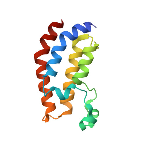 | |
UniProt & NIH Common Fund Data Resources | |||||
PHAROS: Q09472 GTEx: ENSG00000100393 | |||||
Entity Groups | |||||
| Sequence Clusters | 30% Identity50% Identity70% Identity90% Identity95% Identity100% Identity | ||||
| UniProt Group | Q09472 | ||||
Sequence AnnotationsExpand | |||||
Reference Sequence | |||||
| Ligands 3 Unique | |||||
|---|---|---|---|---|---|
| ID | Chains | Name / Formula / InChI Key | 2D Diagram | 3D Interactions | |
| N6I (Subject of Investigation/LOI) Download:Ideal Coordinates CCD File | C [auth A], L [auth B] | (3M)-4-{[(2S)-2-(3-chlorophenyl)-2-hydroxyethyl]amino}-3-[4-methyl-6-(morpholin-4-yl)-1H-benzimidazol-2-yl]pyridin-2(1H)-one C25 H26 Cl N5 O3 ZWVZORIKUNOTCS-OAQYLSRUSA-N |  | ||
| PE5 Download:Ideal Coordinates CCD File | D [auth A] | 3,6,9,12,15,18,21,24-OCTAOXAHEXACOSAN-1-OL C18 H38 O9 CUDPPTPIUWYGFI-UHFFFAOYSA-N |  | ||
| EDO Download:Ideal Coordinates CCD File | E [auth A] F [auth A] G [auth A] H [auth A] I [auth A] | 1,2-ETHANEDIOL C2 H6 O2 LYCAIKOWRPUZTN-UHFFFAOYSA-N |  | ||
| Length ( Å ) | Angle ( ˚ ) |
|---|---|
| a = 43.23 | α = 90 |
| b = 43.23 | β = 90 |
| c = 270.89 | γ = 120 |
| Software Name | Purpose |
|---|---|
| PHENIX | refinement |
| PDB_EXTRACT | data extraction |
| XDS | data reduction |
| XSCALE | data scaling |
| PHENIX | phasing |
| Funding Organization | Location | Grant Number |
|---|---|---|
| Not funded | -- |


