7UAP | pdb_00007uap

Structure of the SARS-CoV-2 S 6P trimer in complex with the neutralizing antibody Fab fragment, C1520


Experimental Data Snapshot

  • Method: ELECTRON MICROSCOPY
  • Resolution: 2.80 Å
  • Aggregation State: PARTICLE 
  • Reconstruction Method: SINGLE PARTICLE 

Starting Model: experimental
View more details

wwPDB Validation 3D Report Full Report

Validation slider image for 7UAP

This is version 1.3 of the entry. See complete history

Literature

Analysis of memory B cells identifies conserved neutralizing epitopes on the N-terminal domain of variant SARS-Cov-2 spike proteins.

Wang, Z.Muecksch, F.Cho, A.Gaebler, C.Hoffmann, H.H.Ramos, V.Zong, S.Cipolla, M.Johnson, B.Schmidt, F.DaSilva, J.Bednarski, E.Ben Tanfous, T.Raspe, R.Yao, K.Lee, Y.E.Chen, T.Turroja, M.Milard, K.G.Dizon, J.Kaczynska, A.Gazumyan, A.Oliveira, T.Y.Rice, C.M.Caskey, M.Bieniasz, P.D.Hatziioannou, T.Barnes, C.O.Nussenzweig, M.C.

(2022) Immunity 55: 998-1012.e8

  • DOI: https://doi.org/10.1016/j.immuni.2022.04.003
  • Primary Citation Related Structures: 
    7UAP, 7UAQ, 7UAR

  • PubMed Abstract: 

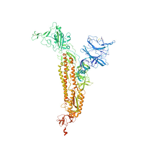
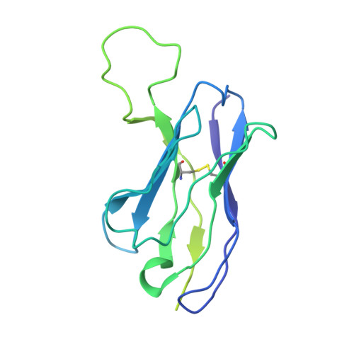
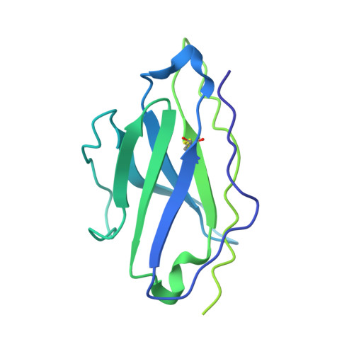
    SARS-CoV-2 infection or vaccination produces neutralizing antibody responses that contribute to better clinical outcomes. The receptor-binding domain (RBD) and the N-terminal domain (NTD) of the spike trimer (S) constitute the two major neutralizing targets for antibodies. Here, we use NTD-specific probes to capture anti-NTD memory B cells in a longitudinal cohort of infected individuals, some of whom were vaccinated. We found 6 complementation groups of neutralizing antibodies. 58% targeted epitopes outside the NTD supersite, 58% neutralized either Gamma or Omicron, and 14% were broad neutralizers that also neutralized Omicron. Structural characterization revealed that broadly active antibodies targeted three epitopes outside the NTD supersite including a class that recognized both the NTD and SD2 domain. Rapid recruitment of memory B cells producing these antibodies into the plasma cell compartment upon re-infection likely contributes to the relatively benign course of subsequent infections with SARS-CoV-2 variants, including Omicron.


  • Organizational Affiliation
    • Laboratory of Molecular Immunology, The Rockefeller University, New York, NY 10065, USA.

Macromolecule Content 

  • Total Structure Weight: 575.74 kDa 
  • Atom Count: 31,657 
  • Modeled Residue Count: 3,957 
  • Deposited Residue Count: 5,118 
  • Unique protein chains: 3

Macromolecules

Find similar proteins by:|  3D Structure
Entity ID: 1
MoleculeChains  Sequence LengthOrganismDetailsImage
Spike glycoprotein
A, B, C
1,256Severe acute respiratory syndrome coronavirus 2Mutation(s): 0 
Gene Names: S2
UniProt
Find proteins for P0DTC2 (Severe acute respiratory syndrome coronavirus 2)
Explore P0DTC2 
Go to UniProtKB:  P0DTC2
Entity Groups
Sequence Clusters30% Identity50% Identity70% Identity90% Identity95% Identity100% Identity
UniProt GroupP0DTC2
Glycosylation
Glycosylation Sites: 15Go to GlyGen: P0DTC2-1
Sequence Annotations
Expand
Reference Sequence
Find similar proteins by:|  3D Structure
Entity ID: 2
MoleculeChains  Sequence LengthOrganismDetailsImage
C1520 Fab Heavy ChainD [auth H],
F [auth M],
H [auth O]
233Homo sapiensMutation(s): 0 
Find similar proteins by:|  3D Structure
Entity ID: 3
MoleculeChains  Sequence LengthOrganismDetailsImage
C1520 Fab Light ChainE [auth L],
G [auth N],
I [auth P]
217Homo sapiensMutation(s): 0 

Oligosaccharides

Help  
Entity ID: 4
MoleculeChains Length2D Diagram GlycosylationD Interactions
2-acetamido-2-deoxy-beta-D-glucopyranose-(1-4)-2-acetamido-2-deoxy-beta-D-glucopyranose
J [auth D],
K [auth E],
L [auth F],
M [auth G],
N [auth I],
J [auth D],
K [auth E],
L [auth F],
M [auth G],
N [auth I],
O [auth J],
P [auth K],
Q,
R,
S,
T,
U
2N-Glycosylation
Glycosylation Resources
GlyTouCan: G42666HT
GlyCosmos: G42666HT
GlyGen: G42666HT

Small Molecules

Ligands 1 Unique
IDChains Name / Formula / InChI Key2D Diagram3D Interactions
NAG

Query on NAG


Download:Ideal Coordinates CCD File
AA [auth A]
AB [auth C]
BA [auth A]
BB [auth L]
CA [auth A]
AA [auth A],
AB [auth C],
BA [auth A],
BB [auth L],
CA [auth A],
CB [auth N],
DA [auth A],
DB [auth P],
EA [auth A],
FA [auth A],
GA [auth B],
HA [auth B],
IA [auth B],
JA [auth B],
KA [auth B],
LA [auth B],
MA [auth B],
NA [auth B],
OA [auth B],
PA [auth B],
QA [auth C],
RA [auth C],
SA [auth C],
TA [auth C],
UA [auth C],
V [auth A],
VA [auth C],
W [auth A],
WA [auth C],
X [auth A],
XA [auth C],
Y [auth A],
YA [auth C],
Z [auth A],
ZA [auth C]
2-acetamido-2-deoxy-beta-D-glucopyranose
C8 H15 N O6
OVRNDRQMDRJTHS-FMDGEEDCSA-N

Experimental Data & Validation

Experimental Data

  • Method: ELECTRON MICROSCOPY
  • Resolution: 2.80 Å
  • Aggregation State: PARTICLE 
  • Reconstruction Method: SINGLE PARTICLE 
EM Software:
TaskSoftware PackageVersion
RECONSTRUCTIONcryoSPARC3.3
MODEL REFINEMENTPHENIX1.19

Structure Validation

View Full Validation Report


Entry History 

& Funding Information

Deposition Data

  • Released Date: 2022-04-27 
  • Deposition Author(s): Barnes, C.O.

Funding OrganizationLocationGrant Number
Howard Hughes Medical Institute (HHMI)United StatesGT15335

Revision History  (Full details and data files)

  • Version 1.0: 2022-04-27
    Type: Initial release
  • Version 1.1: 2022-05-04
    Changes: Database references
  • Version 1.2: 2022-06-22
    Changes: Database references
  • Version 1.3: 2024-11-13
    Changes: Data collection, Refinement description, Structure summary