7U2P | pdb_00007u2p

Structure of TcdA GTD in complex with RhoA


Experimental Data Snapshot

  • Method: X-RAY DIFFRACTION
  • Resolution: 2.60 Å
  • R-Value Free: 
    0.235 (Depositor), 0.236 (DCC) 
  • R-Value Work: 
    0.215 (Depositor), 0.216 (DCC) 

Starting Models: experimental
View more details

wwPDB Validation 3D Report Full Report

Validation slider image for 7U2P

Ligand Structure Quality Assessment 


This is version 1.3 of the entry. See complete history

Literature

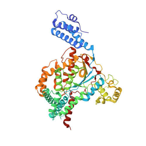
Structure of the glucosyltransferase domain of TcdA in complex with RhoA provides insights into substrate recognition.

Chen, B.Liu, Z.Perry, K.Jin, R.

(2022) Sci Rep 12: 9028-9028

  • DOI: https://doi.org/10.1038/s41598-022-12909-8
  • Primary Citation Related Structures: 
    7U2P

  • PubMed Abstract: 

    Clostridioides difficile is one of the most common causes of antibiotic-associated diarrhea in developed countries. As key virulence factors of C. difficile, toxin A (TcdA) and toxin B (TcdB) act by glucosylating and inactivating Rho and Ras family small GTPases in host cells, which leads to actin cytoskeleton disruption, cell rounding, and ultimately cell death. Here we present the co-crystal structure of the glucosyltransferase domain (GTD) of TcdA in complex with its substrate human RhoA at 2.60-angstrom resolution. This structure reveals that TcdA GTD grips RhoA mainly through its switch I and switch II regions, which is complemented by interactions involving RhoA's pre-switch I region. Comprehensive structural comparisons between the TcdA GTD-RhoA complex and the structures of TcdB GTD in complex with Cdc42 and R-Ras reveal both the conserved and divergent features of these two toxins in terms of substrate recognition. Taken together, these findings establish the structural basis for TcdA recognition of small GTPases and advance our understanding of the substrates selectivity of large clostridial toxins.


  • Organizational Affiliation
    • Department of Physiology and Biophysics, School of Medicine, University of California, Irvine, CA, 92697, USA.

Macromolecule Content 

  • Total Structure Weight: 85 kDa 
  • Atom Count: 5,933 
  • Modeled Residue Count: 721 
  • Deposited Residue Count: 729 
  • Unique protein chains: 2

Macromolecules

Find similar proteins by:|  3D Structure
Entity ID: 1
MoleculeChains  Sequence LengthOrganismDetailsImage
Glucosyltransferase TcdA543Clostridioides difficileMutation(s): 1 
Gene Names: tcdAtoxA
EC: 2.4.1 (PDB Primary Data), 3.4.22 (UniProt)
UniProt
Find proteins for P16154 (Clostridioides difficile)
Explore P16154 
Go to UniProtKB:  P16154
Entity Groups
Sequence Clusters30% Identity50% Identity70% Identity90% Identity95% Identity100% Identity
UniProt GroupP16154
Sequence Annotations
Expand
Reference Sequence
Find similar proteins by:|  3D Structure
Entity ID: 2
MoleculeChains  Sequence LengthOrganismDetailsImage
Transforming protein RhoA186Homo sapiensMutation(s): 1 
Gene Names: RhoaArhaArha2
EC: 3.6.5.2
UniProt & NIH Common Fund Data Resources
Find proteins for P61586 (Homo sapiens)
Explore P61586 
Go to UniProtKB:  P61586
PHAROS:  P61586
GTEx:  ENSG00000067560 
Entity Groups
Sequence Clusters30% Identity50% Identity70% Identity90% Identity95% Identity100% Identity
UniProt GroupP61586
Sequence Annotations
Expand
Reference Sequence

Small Molecules

Ligands 5 Unique
IDChains Name / Formula / InChI Key2D Diagram3D Interactions
UPG
(Subject of Investigation/LOI)

Query on UPG


Download:Ideal Coordinates CCD File
C [auth A]URIDINE-5'-DIPHOSPHATE-GLUCOSE
C15 H24 N2 O17 P2
HSCJRCZFDFQWRP-JZMIEXBBSA-N
GDP
(Subject of Investigation/LOI)

Query on GDP


Download:Ideal Coordinates CCD File
F [auth B]GUANOSINE-5'-DIPHOSPHATE
C10 H15 N5 O11 P2
QGWNDRXFNXRZMB-UUOKFMHZSA-N
SO4
(Subject of Investigation/LOI)

Query on SO4


Download:Ideal Coordinates CCD File
E [auth A]SULFATE ION
O4 S
QAOWNCQODCNURD-UHFFFAOYSA-L
MN
(Subject of Investigation/LOI)

Query on MN


Download:Ideal Coordinates CCD File
D [auth A]MANGANESE (II) ION
Mn
WAEMQWOKJMHJLA-UHFFFAOYSA-N
MG
(Subject of Investigation/LOI)

Query on MG


Download:Ideal Coordinates CCD File
G [auth B]MAGNESIUM ION
Mg
JLVVSXFLKOJNIY-UHFFFAOYSA-N

Experimental Data & Validation

Experimental Data

  • Method: X-RAY DIFFRACTION
  • Resolution: 2.60 Å
  • R-Value Free:  0.235 (Depositor), 0.236 (DCC) 
  • R-Value Work:  0.215 (Depositor), 0.216 (DCC) 
Space Group: C 2 2 21
Unit Cell:
Length ( Å )Angle ( ˚ )
a = 114.641α = 90
b = 122.798β = 90
c = 127.474γ = 90
Software Package:
Software NamePurpose
XDSdata reduction
XDSdata scaling
PHENIXphasing
REFMACrefinement

Structure Validation

View Full Validation Report


Ligand Structure Quality Assessment 


Entry History 

& Funding Information

Deposition Data


Funding OrganizationLocationGrant Number
National Institutes of Health/National Institute Of Allergy and Infectious Diseases (NIH/NIAID)United StatesR01AI125704
National Institutes of Health/National Institute Of Allergy and Infectious Diseases (NIH/NIAID)United StatesR01AI139087
National Institutes of Health/National Institute Of Allergy and Infectious Diseases (NIH/NIAID)United StatesR01AI158503
National Institutes of Health/National Institute Of Allergy and Infectious Diseases (NIH/NIAID)United StatesR21AI156092
National Institutes of Health/National Institute of General Medical Sciences (NIH/NIGMS)United StatesP30 GM124165

Revision History  (Full details and data files)

  • Version 1.0: 2022-05-04
    Type: Initial release
  • Version 1.1: 2022-05-11
    Changes: Structure summary
  • Version 1.2: 2022-06-08
    Changes: Database references
  • Version 1.3: 2023-10-18
    Changes: Data collection, Derived calculations, Refinement description