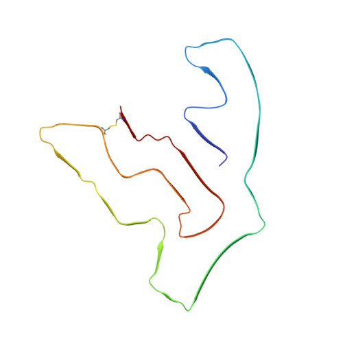
Homotypic fibrillization of TMEM106B across diverse neurodegenerative diseases.
Chang, A., Xiang, X., Wang, J., Lee, C., Arakhamia, T., Simjanoska, M., Wang, C., Carlomagno, Y., Zhang, G., Dhingra, S., Thierry, M., Perneel, J., Heeman, B., Forgrave, L.M., DeTure, M., DeMarco, M.L., Cook, C.N., Rademakers, R., Dickson, D.W., Petrucelli, L., Stowell, M.H.B., Mackenzie, I.R.A., Fitzpatrick, A.W.P.(2022) Cell 185: 1346-1355.e15
- PubMed: 35247328
- DOI: https://doi.org/10.1016/j.cell.2022.02.026
- Primary Citation of Related Structures:
7U0Z, 7U10, 7U11, 7U12, 7U13, 7U14, 7U15, 7U16, 7U17, 7U18 - PubMed Abstract:
Misfolding and aggregation of disease-specific proteins, resulting in the formation of filamentous cellular inclusions, is a hallmark of neurodegenerative disease with characteristic filament structures, or conformers, defining each proteinopathy. Here we show that a previously unsolved amyloid fibril composed of a 135 amino acid C-terminal fragment of TMEM106B is a common finding in distinct human neurodegenerative diseases, including cases characterized by abnormal aggregation of TDP-43, tau, or α-synuclein protein. A combination of cryoelectron microscopy and mass spectrometry was used to solve the structures of TMEM106B fibrils at a resolution of 2.7 Å from postmortem human brain tissue afflicted with frontotemporal lobar degeneration with TDP-43 pathology (FTLD-TDP, n = 8), progressive supranuclear palsy (PSP, n = 2), or dementia with Lewy bodies (DLB, n = 1). The commonality of abundant amyloid fibrils composed of TMEM106B, a lysosomal/endosomal protein, to a broad range of debilitating human disorders indicates a shared fibrillization pathway that may initiate or accelerate neurodegeneration.
- Mortimer B. Zuckerman Mind Brain Behavior Institute, Columbia University, New York, NY 10027, USA; Department of Biochemistry and Molecular Biophysics, Columbia University, New York, NY 10032, USA; Taub Institute for Research on Alzheimer's Disease and the Aging Brain, Columbia University Irving Medical Center, 630 West 168th Street, New York, NY 10032, USA.
Organizational Affiliation:


