7TVG | pdb_00007tvg

Crystal Structure of SHOC2 to a resolution of 2.4 Angstrom


Experimental Data Snapshot

  • Method: X-RAY DIFFRACTION
  • Resolution: 2.40 Å
  • R-Value Free: 
    0.267 (Depositor), 0.267 (DCC) 
  • R-Value Work: 
    0.216 (Depositor), 0.216 (DCC) 
  • R-Value Observed: 
    0.219 (Depositor) 

wwPDB Validation 3D Report Full Report

Validation slider image for 7TVG

This is version 1.4 of the entry. See complete history

Literature

Structure of the SHOC2-MRAS-PP1C complex provides insights into RAF activation and Noonan syndrome.

Bonsor, D.A.Alexander, P.Snead, K.Hartig, N.Drew, M.Messing, S.Finci, L.I.Nissley, D.V.McCormick, F.Esposito, D.Rodriguez-Viciana, P.Stephen, A.G.Simanshu, D.K.

(2022) Nat Struct Mol Biol 29: 966-977

  • DOI: https://doi.org/10.1038/s41594-022-00841-4
  • Primary Citation Related Structures: 
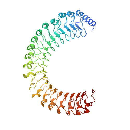
    7TVF, 7TVG

  • PubMed Abstract: 

    SHOC2 acts as a strong synthetic lethal interactor with MEK inhibitors in multiple KRAS cancer cell lines. SHOC2 forms a heterotrimeric complex with MRAS and PP1C that is essential for regulating RAF and MAPK-pathway activation by dephosphorylating a specific phosphoserine on RAF kinases. Here we present the high-resolution crystal structure of the SHOC2-MRAS-PP1C (SMP) complex and apo-SHOC2. Our structures reveal that SHOC2, MRAS, and PP1C form a stable ternary complex in which all three proteins synergistically interact with each other. Our results show that dephosphorylation of RAF substrates by PP1C is enhanced upon interacting with SHOC2 and MRAS. The SMP complex forms only when MRAS is in an active state and is dependent on SHOC2 functioning as a scaffolding protein in the complex by bringing PP1C and MRAS together. Our results provide structural insights into the role of the SMP complex in RAF activation and how mutations found in Noonan syndrome enhance complex formation, and reveal new avenues for therapeutic interventions.


  • Organizational Affiliation
    • NCI RAS Initiative, Cancer Research Technology Program, Frederick National Laboratory for Cancer Research, Frederick, MD, USA.

Macromolecule Content 

  • Total Structure Weight: 57.28 kDa 
  • Atom Count: 3,809 
  • Modeled Residue Count: 480 
  • Deposited Residue Count: 507 
  • Unique protein chains: 1

Macromolecules

Find similar proteins by:|  3D Structure
Entity ID: 1
MoleculeChains  Sequence LengthOrganismDetailsImage
Leucine-rich repeat protein SHOC-2A [auth D]507Homo sapiensMutation(s): 0 
Gene Names: SHOC2KIAA0862
UniProt & NIH Common Fund Data Resources
Find proteins for Q9UQ13 (Homo sapiens)
Explore Q9UQ13 
Go to UniProtKB:  Q9UQ13
PHAROS:  Q9UQ13
GTEx:  ENSG00000108061 
Entity Groups
Sequence Clusters30% Identity50% Identity70% Identity90% Identity95% Identity100% Identity
UniProt GroupQ9UQ13
Sequence Annotations
Expand
Reference Sequence

Experimental Data & Validation

Experimental Data

  • Method: X-RAY DIFFRACTION
  • Resolution: 2.40 Å
  • R-Value Free:  0.267 (Depositor), 0.267 (DCC) 
  • R-Value Work:  0.216 (Depositor), 0.216 (DCC) 
  • R-Value Observed: 0.219 (Depositor) 
Space Group: P 32
Unit Cell:
Length ( Å )Angle ( ˚ )
a = 77.567α = 90
b = 77.567β = 90
c = 83.015γ = 120
Software Package:
Software NamePurpose
PHENIXrefinement
PDB_EXTRACTdata extraction
XDSdata reduction
Aimlessdata scaling
PHASERphasing

Structure Validation

View Full Validation Report


Entry History 

& Funding Information

Deposition Data


Funding OrganizationLocationGrant Number
National Institutes of Health/National Cancer Institute (NIH/NCI)United StatesHHSN261200800001E

Revision History  (Full details and data files)

  • Version 1.0: 2022-05-04
    Type: Initial release
  • Version 1.1: 2022-10-05
    Changes: Database references
  • Version 1.2: 2022-10-12
    Changes: Database references
  • Version 1.3: 2022-10-26
    Changes: Database references
  • Version 1.4: 2024-05-22
    Changes: Data collection