7TUA | pdb_00007tua

Human Synaptotagmin-1 C2B Y312F without Ca2+


Experimental Data Snapshot

  • Method: X-RAY DIFFRACTION
  • Resolution: 1.35 Å
  • R-Value Free: 
    0.199 (Depositor), 0.199 (DCC) 
  • R-Value Work: 
    0.178 (Depositor), 0.178 (DCC) 
  • R-Value Observed: 
    0.179 (Depositor) 

Starting Model: experimental
View more details

wwPDB Validation 3D Report Full Report

Validation slider image for 7TUA

This is version 1.2 of the entry. See complete history

Literature

The AD3 locus of synaptotagmin-1 C2 domains modulates domain stability.

Dominguez, M.J.Bui, A.A.Villarreal, J.Snow, A.Karmakar, S.Harsini, F.M.Rock, P.J.Rice, A.M.Fuson, K.L.Sutton, R.B.

(2024) Biophys J 

  • DOI: https://doi.org/10.1016/j.bpj.2024.11.009
  • Primary Citation Related Structures: 
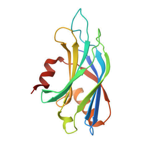
    4WEE, 7TUA, 7TX9, 7U4Q

  • PubMed Abstract: 

    Synaptotagmin-1 (syt1) functions as the Ca 2+ -dependent sensor that triggers the rapid and synchronous release of neurotransmitters from neurotransmitter-containing vesicles during neuronal exocytosis. The syt1 protein has two homologous tandem C2 domains that interact with phospholipids in a Ca 2+ -dependent manner. Despite the crucial role of syt1 in exocytosis, the precise interactions between Ca 2+ , syt1, and phospholipids are not fully understood. In a study involving recessive lethal mutations in the syt1 gene, a specific mutation named AD3 was generated in Drosophila syt1, resulting in a significant reduction in Ca 2+ -dependent exocytosis. Further investigation revealed that the AD3 mutation was a missense mutation located in a conserved consensus sequence within the C2B domain of Drosophila syt1. However, the biophysical impact of the AD3 mutation had not been analyzed. Our study uses x-ray crystallography, isothermal titration calorimetry, thermodynamic analysis, and molecular dynamics simulation to show that the primary defect caused by the AD3 mutation in the syt1 protein is reduced thermodynamic stability. This instability alters the population of Ca 2+ -receptive states, leading to two major consequences: decreased affinity for calcium ions and compromised stabilization of the domain normally enhanced by Ca 2+ . We conclude that this conserved residue acts as a structural constraint, delimiting the movement of loop 3 within the pocket and ultimately influencing the affinity of the calcium ion binding with the C2 domain.


  • Organizational Affiliation
    • Department of Cell Physiology and Molecular Biophysics, Texas Tech University Health Sciences Center, Lubbock, Texas.

Macromolecule Content 

  • Total Structure Weight: 17.96 kDa 
  • Atom Count: 1,484 
  • Modeled Residue Count: 148 
  • Deposited Residue Count: 157 
  • Unique protein chains: 1

Macromolecules

Find similar proteins by:|  3D Structure
Entity ID: 1
MoleculeChains  Sequence LengthOrganismDetailsImage
Synaptotagmin157Homo sapiensMutation(s): 0 
Gene Names: SYT1hCG_2016754
Membrane Entity: Yes 
UniProt & NIH Common Fund Data Resources
Find proteins for P21579 (Homo sapiens)
Explore P21579 
Go to UniProtKB:  P21579
PHAROS:  P21579
GTEx:  ENSG00000067715 
Entity Groups
Sequence Clusters30% Identity50% Identity70% Identity90% Identity95% Identity100% Identity
UniProt GroupP21579
Sequence Annotations
Expand
Reference Sequence

Small Molecules

Ligands 1 Unique
IDChains Name / Formula / InChI Key2D Diagram3D Interactions
SO4

Query on SO4


Download:Ideal Coordinates CCD File
B [auth A]SULFATE ION
O4 S
QAOWNCQODCNURD-UHFFFAOYSA-L

Experimental Data & Validation

Experimental Data

  • Method: X-RAY DIFFRACTION
  • Resolution: 1.35 Å
  • R-Value Free:  0.199 (Depositor), 0.199 (DCC) 
  • R-Value Work:  0.178 (Depositor), 0.178 (DCC) 
  • R-Value Observed: 0.179 (Depositor) 
Space Group: P 32 2 1
Unit Cell:
Length ( Å )Angle ( ˚ )
a = 54.44α = 90
b = 54.44β = 90
c = 104.07γ = 120
Software Package:
Software NamePurpose
PHASERphasing
PHENIXrefinement
PDB-REDOrefinement
xia2data scaling
xia2data reduction
XDSdata reduction
XSCALEdata scaling
Cootmodel building

Structure Validation

View Full Validation Report


Entry History 

& Funding Information

Deposition Data


Funding OrganizationLocationGrant Number
National Institutes of Health/National Institute of Mental Health (NIH/NIMH)United States1R21MH070589-01

Revision History  (Full details and data files)

  • Version 1.0: 2023-03-22
    Type: Initial release
  • Version 1.1: 2023-10-25
    Changes: Data collection, Refinement description
  • Version 1.2: 2025-01-15
    Changes: Database references, Structure summary