7TTR | pdb_00007ttr

Skd3_ATPyS_FITC-casein Hexamer, AAA+ only


Experimental Data Snapshot

  • Method: ELECTRON MICROSCOPY
  • Resolution: 2.96 Å
  • Aggregation State: PARTICLE 
  • Reconstruction Method: SINGLE PARTICLE 

wwPDB Validation 3D Report Full Report

Validation slider image for 7TTR

This is version 1.3 of the entry. See complete history

Literature

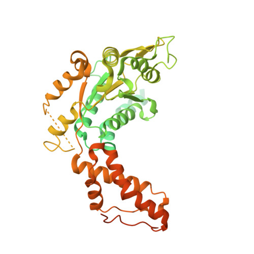
Unique structural features govern the activity of a human mitochondrial AAA+ disaggregase, Skd3.

Cupo, R.R.Rizo, A.N.Braun, G.A.Tse, E.Chuang, E.Gupta, K.Southworth, D.R.Shorter, J.

(2022) Cell Rep 40: 111408-111408

  • DOI: https://doi.org/10.1016/j.celrep.2022.111408
  • Primary Citation Related Structures: 
    7TTR, 7TTS

  • PubMed Abstract: 

    The AAA+ protein, Skd3 (human CLPB), solubilizes proteins in the mitochondrial intermembrane space, which is critical for human health. Skd3 variants with defective protein-disaggregase activity cause severe congenital neutropenia (SCN) and 3-methylglutaconic aciduria type 7 (MGCA7). How Skd3 disaggregates proteins remains poorly understood. Here, we report a high-resolution structure of a Skd3-substrate complex. Skd3 adopts a spiral hexameric arrangement that engages substrate via pore-loop interactions in the nucleotide-binding domain (NBD). Substrate-bound Skd3 hexamers stack head-to-head via unique, adaptable ankyrin-repeat domain (ANK)-mediated interactions to form dodecamers. Deleting the ANK linker region reduces dodecamerization and disaggregase activity. We elucidate apomorphic features of the Skd3 NBD and C-terminal domain that regulate disaggregase activity. We also define how Skd3 subunits collaborate to disaggregate proteins. Importantly, SCN-linked subunits sharply inhibit disaggregase activity, whereas MGCA7-linked subunits do not. These advances illuminate Skd3 structure and mechanism, explain SCN and MGCA7 inheritance patterns, and suggest therapeutic strategies.


  • Organizational Affiliation
    • Department of Biochemistry and Biophysics, University of Pennsylvania, Philadelphia, PA, USA; Pharmacology Graduate Group, Perelman School of Medicine, University of Pennsylvania, Philadelphia, PA, USA.

Macromolecule Content 

  • Total Structure Weight: 424.07 kDa 
  • Atom Count: 15,697 
  • Modeled Residue Count: 1,926 
  • Deposited Residue Count: 3,728 
  • Unique protein chains: 2

Macromolecules

Find similar proteins by:|  3D Structure
Entity ID: 1
MoleculeChains  Sequence LengthOrganismDetailsImage
Caseinolytic peptidase B protein homolog
A, B, C, D, E
A, B, C, D, E, F
584Homo sapiensMutation(s): 0 
Gene Names: CLPBHSP78SKD3
EC: 3.6.1
UniProt & NIH Common Fund Data Resources
Find proteins for Q9H078 (Homo sapiens)
Explore Q9H078 
Go to UniProtKB:  Q9H078
PHAROS:  Q9H078
GTEx:  ENSG00000162129 
Entity Groups
Sequence Clusters30% Identity50% Identity70% Identity90% Identity95% Identity100% Identity
UniProt GroupQ9H078
Sequence Annotations
Expand
Reference Sequence
Find similar proteins by:|  3D Structure
Entity ID: 2
MoleculeChains  Sequence LengthOrganismDetailsImage
Beta-caseinG [auth P]224Bos taurusMutation(s): 0 
UniProt
Find proteins for P02666 (Bos taurus)
Explore P02666 
Go to UniProtKB:  P02666
Entity Groups
Sequence Clusters30% Identity50% Identity70% Identity90% Identity95% Identity100% Identity
UniProt GroupP02666
Sequence Annotations
Expand
Reference Sequence

Small Molecules

Ligands 3 Unique
IDChains Name / Formula / InChI Key2D Diagram3D Interactions
AGS
(Subject of Investigation/LOI)

Query on AGS


Download:Ideal Coordinates CCD File
J [auth B],
L [auth C],
M [auth D],
O [auth E]
PHOSPHOTHIOPHOSPHORIC ACID-ADENYLATE ESTER
C10 H16 N5 O12 P3 S
NLTUCYMLOPLUHL-KQYNXXCUSA-N
ADP
(Subject of Investigation/LOI)

Query on ADP


Download:Ideal Coordinates CCD File
H [auth A]ADENOSINE-5'-DIPHOSPHATE
C10 H15 N5 O10 P2
XTWYTFMLZFPYCI-KQYNXXCUSA-N
MG
(Subject of Investigation/LOI)

Query on MG


Download:Ideal Coordinates CCD File
I [auth B],
K [auth C],
N [auth D],
P [auth E]
MAGNESIUM ION
Mg
JLVVSXFLKOJNIY-UHFFFAOYSA-N

Experimental Data & Validation

Experimental Data

  • Method: ELECTRON MICROSCOPY
  • Resolution: 2.96 Å
  • Aggregation State: PARTICLE 
  • Reconstruction Method: SINGLE PARTICLE 
EM Software:
TaskSoftware PackageVersion
MODEL REFINEMENTPHENIX

Structure Validation

View Full Validation Report


Entry History 

& Funding Information

Deposition Data

  • Released Date: 2022-09-28 
  • Deposition Author(s): Rizo, A.N.

Funding OrganizationLocationGrant Number
National Institutes of Health/National Institute of General Medical Sciences (NIH/NIGMS)United StatesR01GM138690
National Institutes of Health/National Institute of General Medical Sciences (NIH/NIGMS)United StatesR01GM099836

Revision History  (Full details and data files)

  • Version 1.0: 2022-09-28
    Type: Initial release
  • Version 1.1: 2022-10-12
    Changes: Database references
  • Version 1.2: 2024-06-12
    Changes: Data collection
  • Version 1.3: 2025-05-14
    Changes: Data collection, Structure summary