7TR1 | pdb_00007tr1

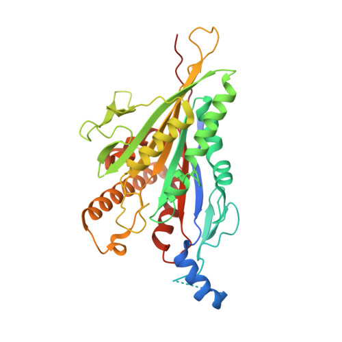
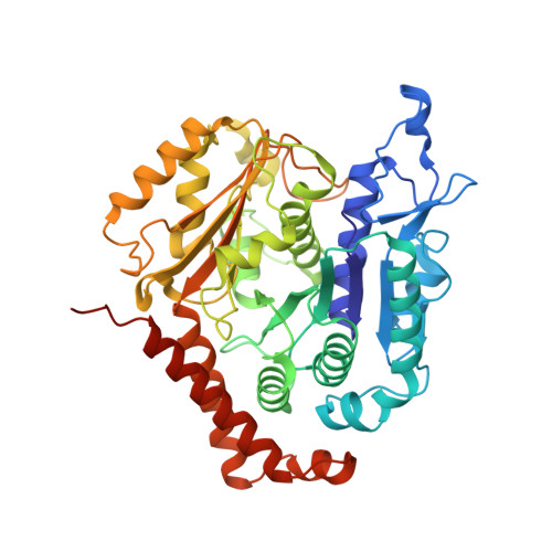
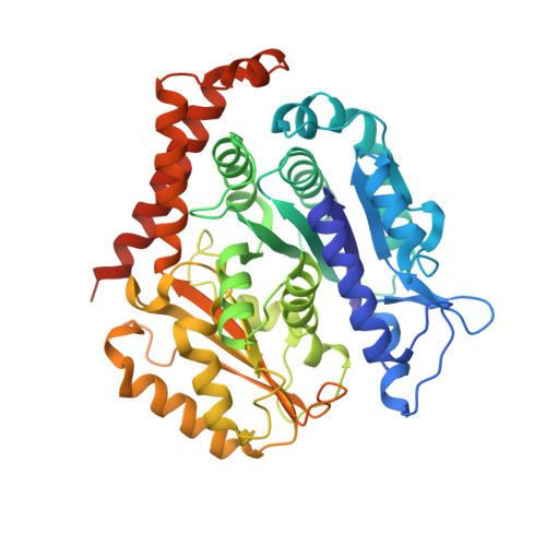
CaKip3[2-436]-L2-mutant(HsKHC) - AMP-PNP in complex with a microtubule


Experimental Data Snapshot

  • Method: ELECTRON MICROSCOPY
  • Resolution: 3.10 Å
  • Aggregation State: FILAMENT 
  • Reconstruction Method: HELICAL 

wwPDB Validation 3D Report Full Report

Validation slider image for 7TR1

This is version 1.2 of the entry. See complete history. 

Literature

Kinesin-8-specific loop-2 controls the dual activities of the motor domain according to tubulin protofilament shape.

Hunter, B., Benoit, M.P.M.H., Asenjo, A.B., Doubleday, C., Trofimova, D., Frazer, C., Shoukat, I., Sosa, H., Allingham, J.S.

(2022) Nat Commun 13: 4198-4198

  • DOI: https://doi.org/10.1038/s41467-022-31794-3
  • Primary Citation Related Structures: 
    7LFF, 7TQX, 7TQY, 7TQZ, 7TR0, 7TR1, 7TR2, 7TR3

  • PubMed Abstract: 

    Kinesin-8s are dual-activity motor proteins that can move processively on microtubules and depolymerize microtubule plus-ends, but their mechanism of combining these distinct activities remains unclear. We addressed this by obtaining cryo-EM structures (2.6-3.9 Å) of Candida albicans Kip3 in different catalytic states on the microtubule lattice and on a curved microtubule end mimic. We also determined a crystal structure of microtubule-unbound CaKip3-ADP (2.0 Å) and analyzed the biochemical activity of CaKip3 and kinesin-1 mutants. These data reveal that the microtubule depolymerization activity of kinesin-8 originates from conformational changes of its motor core that are amplified by dynamic contacts between its extended loop-2 and tubulin. On curved microtubule ends, loop-1 inserts into preceding motor domains, forming head-to-tail arrays of kinesin-8s that complement loop-2 contacts with curved tubulin and assist depolymerization. On straight tubulin protofilaments in the microtubule lattice, loop-2-tubulin contacts inhibit conformational changes in the motor core, but in the ADP-Pi state these contacts are relaxed, allowing neck-linker docking for motility. We propose that these tubulin shape-induced alternations between pro-microtubule-depolymerization and pro-motility kinesin states, regulated by loop-2, are the key to the dual activity of kinesin-8 motors.


  • Organizational Affiliation: 
    • Department of Biomedical and Molecular Sciences, Queen's University, Kingston, ON, K7L 3N6, Canada.

Macromolecule Content 

  • Total Structure Weight: 148.92 kDa 
  • Atom Count: 9,707 
  • Modeled Residue Count: 1,216 
  • Deposited Residue Count: 1,316 
  • Unique protein chains: 3

Macromolecules

Find similar proteins by:|  3D Structure
Entity ID: 1
MoleculeChains  Sequence LengthOrganismDetailsImage
Kinesin-like protein,Kinesin-1 heavy chainA [auth K]420Candida albicans, Homo sapiensMutation(s): 0 
Gene Names: CAWG_02882, KIF5B, KNS, KNS1
UniProt & NIH Common Fund Data Resources
Find proteins for C4YNU9 (Candida albicans (strain WO-1))
Explore C4YNU9 
Go to UniProtKB:  C4YNU9
Find proteins for P33176 (Homo sapiens)
Explore P33176 
Go to UniProtKB:  P33176
PHAROS:  P33176
GTEx:  ENSG00000170759 
Entity Groups
Sequence Clusters30% Identity50% Identity70% Identity90% Identity95% Identity100% Identity
UniProt GroupsP33176C4YNU9
Sequence Annotations
Expand
Reference Sequence
Find similar proteins by:|  3D Structure
Entity ID: 2
MoleculeChains  Sequence LengthOrganismDetailsImage
Tubulin alpha-1B chainB [auth A]451Sus scrofaMutation(s): 0 
EC: 3.6.5
UniProt
Find proteins for Q2XVP4 (Sus scrofa)
Explore Q2XVP4 
Go to UniProtKB:  Q2XVP4
Entity Groups
Sequence Clusters30% Identity50% Identity70% Identity90% Identity95% Identity100% Identity
UniProt GroupQ2XVP4
Sequence Annotations
Expand
Reference Sequence
Find similar proteins by:|  3D Structure
Entity ID: 3
MoleculeChains  Sequence LengthOrganismDetailsImage
Tubulin beta-2B chainC [auth B]445Sus scrofaMutation(s): 0 
UniProt
Find proteins for P02554 (Sus scrofa)
Explore P02554 
Go to UniProtKB:  P02554
Entity Groups
Sequence Clusters30% Identity50% Identity70% Identity90% Identity95% Identity100% Identity
UniProt GroupP02554
Sequence Annotations
Expand
Reference Sequence

Small Molecules

Ligands 5 Unique
IDChains Name / Formula / InChI Key2D Diagram3D Interactions
TA1

Query on TA1


Download:Ideal Coordinates CCD File
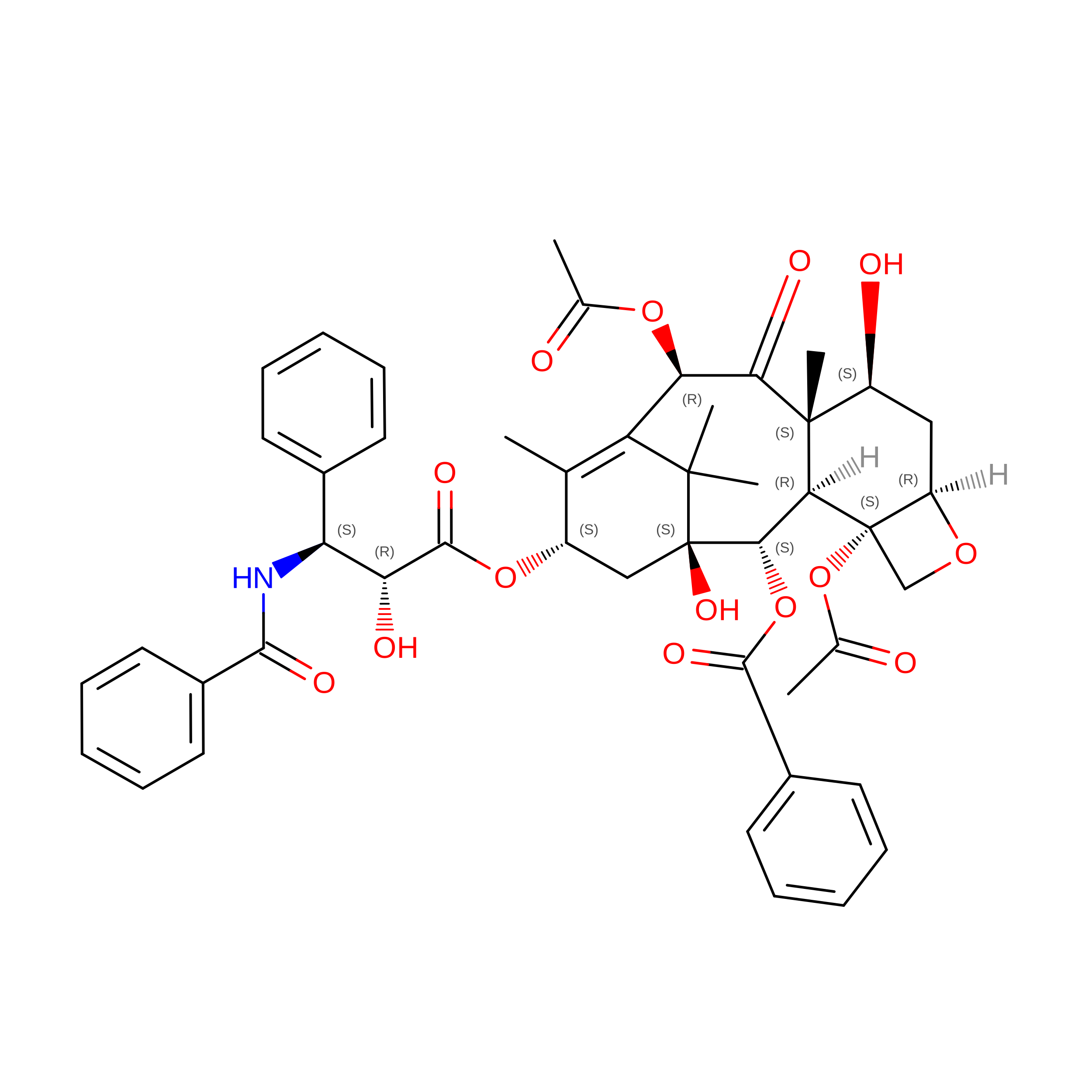
I [auth B]TAXOL
C47 H51 N O14
RCINICONZNJXQF-MZXODVADSA-N
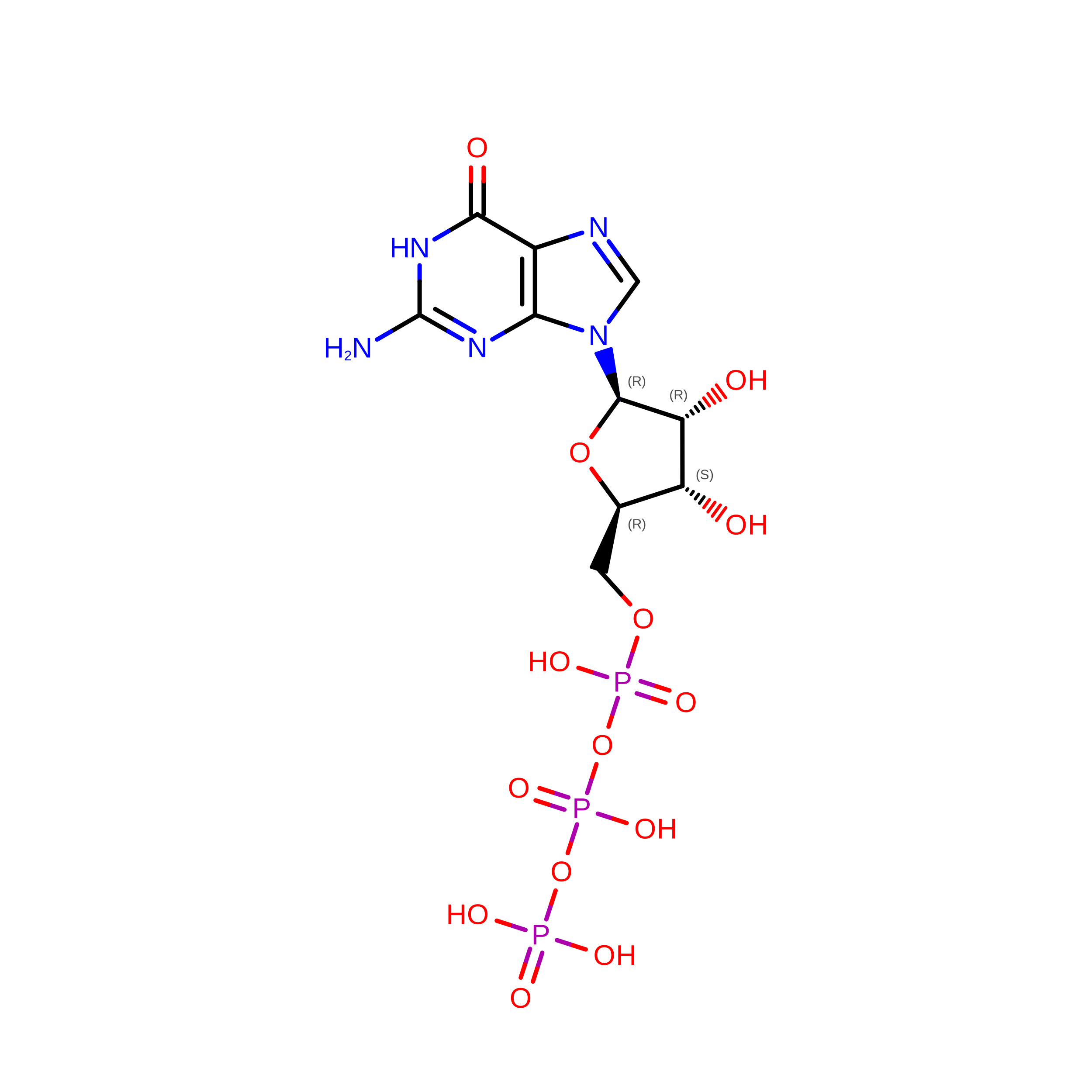
GTP

Query on GTP


Download:Ideal Coordinates CCD File
F [auth A]GUANOSINE-5'-TRIPHOSPHATE
C10 H16 N5 O14 P3
XKMLYUALXHKNFT-UUOKFMHZSA-N
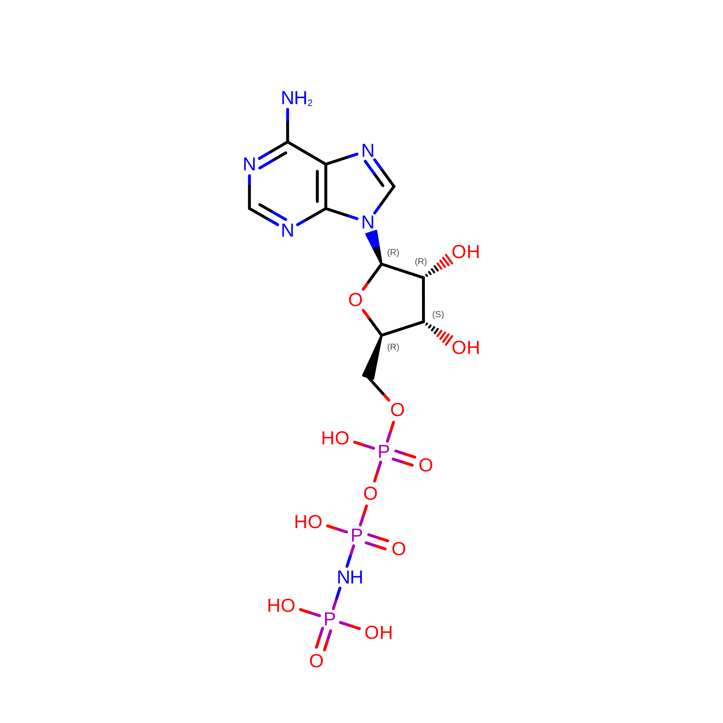
ANP

Query on ANP


Download:Ideal Coordinates CCD File
E [auth K]PHOSPHOAMINOPHOSPHONIC ACID-ADENYLATE ESTER
C10 H17 N6 O12 P3
PVKSNHVPLWYQGJ-KQYNXXCUSA-N
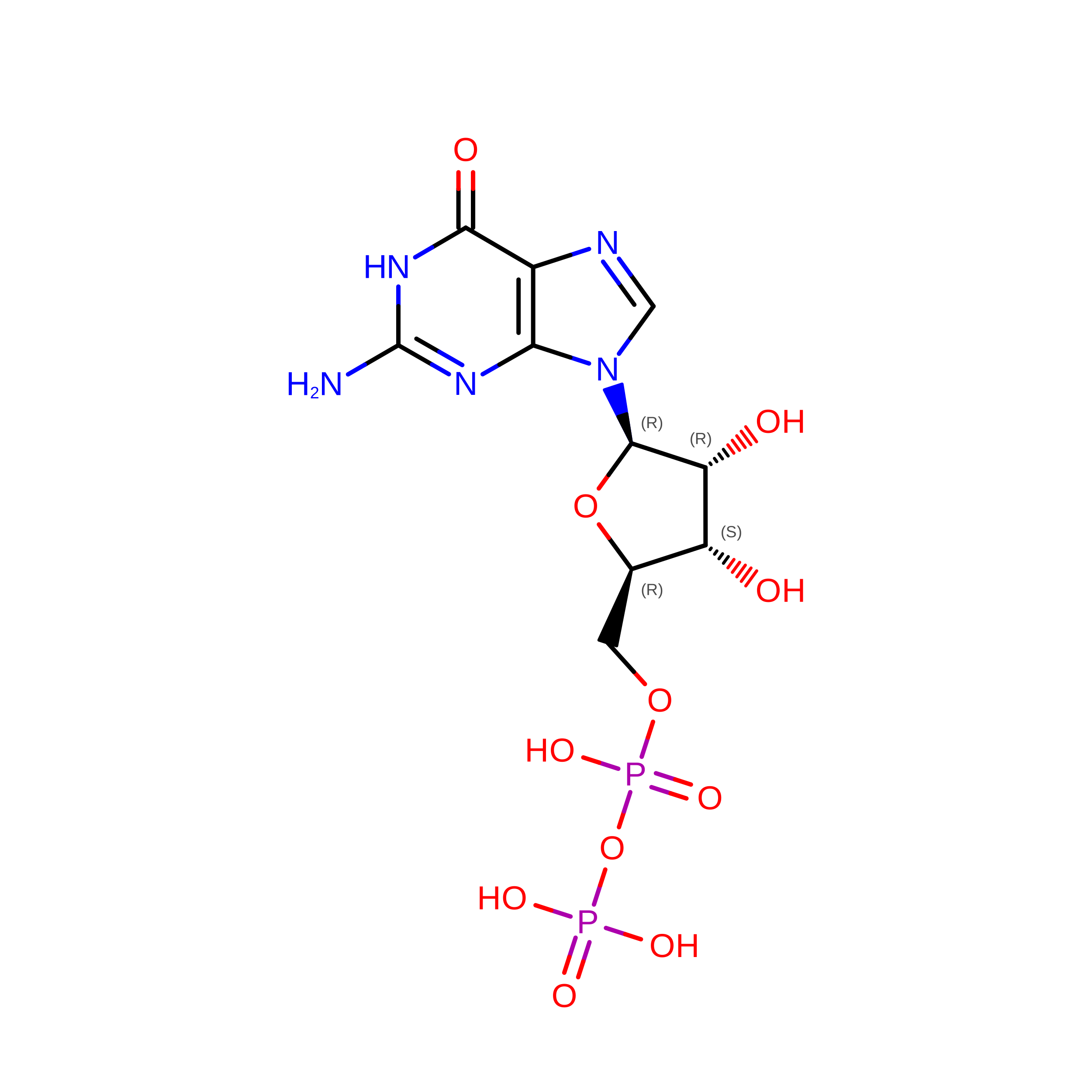
GDP

Query on GDP


Download:Ideal Coordinates CCD File
H [auth B]GUANOSINE-5'-DIPHOSPHATE
C10 H15 N5 O11 P2
QGWNDRXFNXRZMB-UUOKFMHZSA-N
MG

Query on MG


Download:Ideal Coordinates CCD File
D [auth K],
G [auth A]
MAGNESIUM ION
Mg
JLVVSXFLKOJNIY-UHFFFAOYSA-N

Experimental Data & Validation

Experimental Data

  • Method: ELECTRON MICROSCOPY
  • Resolution: 3.10 Å
  • Aggregation State: FILAMENT 
  • Reconstruction Method: HELICAL 
EM Software:
TaskSoftware PackageVersion
RECONSTRUCTIONRELION3.1

Structure Validation

View Full Validation Report


Entry History 

& Funding Information

Deposition Data


Funding OrganizationLocationGrant Number
National Institutes of Health/National Institute of General Medical Sciences (NIH/NIGMS)United StatesR01GM113164
Natural Sciences and Engineering Research Council (NSERC, Canada)CanadaRGPIN/356025-2013
Canadian Institutes of Health Research (CIHR)CanadaMOP-97832

Revision History  (Full details and data files)

  • Version 1.0: 2022-07-20
    Type: Initial release
  • Version 1.1: 2022-08-10
    Changes: Database references
  • Version 1.2: 2024-02-21
    Changes: Data collection