7TFS | pdb_00007tfs

Cryo-EM of the OmcE nanowires from Geobacter sulfurreducens


Experimental Data Snapshot

  • Method: ELECTRON MICROSCOPY
  • Resolution: 4.30 Å
  • Aggregation State: FILAMENT 
  • Reconstruction Method: HELICAL 

wwPDB Validation 3D Report Full Report

Validation slider image for 7TFS

This is version 1.3 of the entry. See complete history

Literature

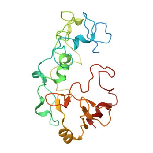
Cryo-EM structure of an extracellular Geobacter OmcE cytochrome filament reveals tetrahaem packing.

Wang, F.Mustafa, K.Suciu, V.Joshi, K.Chan, C.H.Choi, S.Su, Z.Si, D.Hochbaum, A.I.Egelman, E.H.Bond, D.R.

(2022) Nat Microbiol 7: 1291-1300

  • DOI: https://doi.org/10.1038/s41564-022-01159-z
  • Primary Citation Related Structures: 
    7TFS, 7TGG

  • PubMed Abstract: 

    Electrically conductive appendages from the anaerobic bacterium Geobacter sulfurreducens were first observed two decades ago, with genetic and biochemical data suggesting that conductive fibres were type IV pili. Recently, an extracellular conductive filament of G. sulfurreducens was found to contain polymerized c-type cytochrome OmcS subunits, not pilin subunits. Here we report that G. sulfurreducens also produces a second, thinner appendage comprised of cytochrome OmcE subunits and solve its structure using cryo-electron microscopy at ~4.3 Å resolution. Although OmcE and OmcS subunits have no overall sequence or structural similarities, upon polymerization both form filaments that share a conserved haem packing arrangement in which haems are coordinated by histidines in adjacent subunits. Unlike OmcS filaments, OmcE filaments are highly glycosylated. In extracellular fractions from G. sulfurreducens, we detected type IV pili comprising PilA-N and -C chains, along with abundant B-DNA. OmcE is the second cytochrome filament to be characterized using structural and biophysical methods. We propose that there is a broad class of conductive bacterial appendages with conserved haem packing (rather than sequence homology) that enable long-distance electron transport to chemicals or other microbial cells.


  • Organizational Affiliation
    • Department of Biochemistry and Molecular Genetics, University of Virginia School of Medicine, Charlottesville, VA, USA.

Macromolecule Content 

  • Total Structure Weight: 26.31 kDa 
  • Atom Count: 1,626 
  • Modeled Residue Count: 202 
  • Deposited Residue Count: 232 
  • Unique protein chains: 1

Macromolecules

Find similar proteins by:|  3D Structure
Entity ID: 1
MoleculeChains  Sequence LengthOrganismDetailsImage
Cytochrome c232Geobacter sulfurreducensMutation(s): 0 
UniProt
Find proteins for Q74FJ0 (Geobacter sulfurreducens (strain ATCC 51573 / DSM 12127 / PCA))
Explore Q74FJ0 
Go to UniProtKB:  Q74FJ0
Entity Groups
Sequence Clusters30% Identity50% Identity70% Identity90% Identity95% Identity100% Identity
UniProt GroupQ74FJ0
Sequence Annotations
Expand
Reference Sequence

Experimental Data & Validation

Experimental Data

  • Method: ELECTRON MICROSCOPY
  • Resolution: 4.30 Å
  • Aggregation State: FILAMENT 
  • Reconstruction Method: HELICAL 
EM Software:
TaskSoftware PackageVersion
MODEL REFINEMENTPHENIX

Structure Validation

View Full Validation Report


Entry History 

& Funding Information

Deposition Data


Funding OrganizationLocationGrant Number
National Institutes of Health/National Institute of General Medical Sciences (NIH/NIGMS)United StatesR35GM122510
National Institutes of Health/National Institute of General Medical Sciences (NIH/NIGMS)United StatesK99GM138756

Revision History  (Full details and data files)

  • Version 1.0: 2022-05-04
    Type: Initial release
  • Version 1.1: 2022-11-16
    Changes: Database references
  • Version 1.2: 2024-12-25
    Changes: Advisory, Data collection, Derived calculations, Structure summary
  • Version 1.3: 2025-05-14
    Changes: Data collection