7TCY | pdb_00007tcy

The ubiquitin-associated domain of human thirty-eight negative kinase I


Experimental Data Snapshot

  • Method: X-RAY DIFFRACTION
  • Resolution: 1.54 Å
  • R-Value Free: 
    0.214 (Depositor), 0.215 (DCC) 
  • R-Value Work: 
    0.189 (Depositor), 0.190 (DCC) 
  • R-Value Observed: 
    0.190 (Depositor) 

wwPDB Validation 3D Report Full Report

Validation slider image for 7TCY

This is version 1.2 of the entry. See complete history

Literature

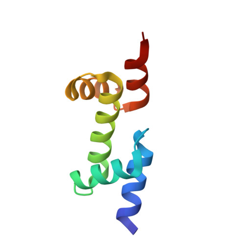
Fusion crystallization reveals the behavior of both the 1TEL crystallization chaperone and the TNK1 UBA domain.

Nawarathnage, S.Tseng, Y.J.Soleimani, S.Smith, T.Pedroza Romo, M.J.Abiodun, W.O.Egbert, C.M.Madhusanka, D.Bunn, D.Woods, B.Tsubaki, E.Stewart, C.Brown, S.Doukov, T.Andersen, J.L.Moody, J.D.

(2023) Structure 31: 1589-1603.e6

  • DOI: https://doi.org/10.1016/j.str.2023.09.001
  • Primary Citation Related Structures: 
    7T8J, 7TCY, 7TDY, 7U4W, 7U4Z

  • PubMed Abstract: 

    Human thirty-eight-negative kinase-1 (TNK1) is implicated in cancer progression. The TNK1 ubiquitin-associated (UBA) domain binds polyubiquitin and plays a regulatory role in TNK1 activity and stability. No experimentally determined molecular structure of this unusual UBA domain is available. We fused the UBA domain to the 1TEL variant of the translocation ETS leukemia protein sterile alpha motif (TELSAM) crystallization chaperone and obtained crystals diffracting as far as 1.53 Å. GG and GSGG linkers allowed the UBA to reproducibly find a productive binding mode against its host 1TEL polymer and crystallize at protein concentrations as low as 0.2 mg/mL. Our studies support a mechanism of 1TEL fusion crystallization and show that 1TEL fusion crystals require fewer crystal contacts than traditional protein crystals. Modeling and experimental validation suggest the UBA domain may be selective for both the length and linkages of polyubiquitin chains.


  • Organizational Affiliation
    • Department of Chemistry and Biochemistry, Brigham Young University, Provo, UT, USA.

Macromolecule Content 

  • Total Structure Weight: 17.88 kDa 
  • Atom Count: 1,382 
  • Modeled Residue Count: 154 
  • Deposited Residue Count: 156 
  • Unique protein chains: 1

Macromolecules

Find similar proteins by:|  3D Structure
Entity ID: 1
MoleculeChains  Sequence LengthOrganismDetailsImage
Non-receptor tyrosine-protein kinase TNK1
A, B
78Homo sapiensMutation(s): 3 
Gene Names: TNK1
EC: 2.7.10.2
UniProt & NIH Common Fund Data Resources
Find proteins for Q13470 (Homo sapiens)
Explore Q13470 
Go to UniProtKB:  Q13470
PHAROS:  Q13470
GTEx:  ENSG00000174292 
Entity Groups
Sequence Clusters30% Identity50% Identity70% Identity90% Identity95% Identity100% Identity
UniProt GroupQ13470
Sequence Annotations
Expand
Reference Sequence

Small Molecules

Ligands 4 Unique
IDChains Name / Formula / InChI Key2D Diagram3D Interactions
PO4

Query on PO4


Download:Ideal Coordinates CCD File
D [auth A]PHOSPHATE ION
O4 P
NBIIXXVUZAFLBC-UHFFFAOYSA-K
FMT

Query on FMT


Download:Ideal Coordinates CCD File
C [auth A],
F [auth A],
I [auth A],
J [auth B]
FORMIC ACID
C H2 O2
BDAGIHXWWSANSR-UHFFFAOYSA-N
CL

Query on CL


Download:Ideal Coordinates CCD File
G [auth A],
H [auth A]
CHLORIDE ION
Cl
VEXZGXHMUGYJMC-UHFFFAOYSA-M
MG

Query on MG


Download:Ideal Coordinates CCD File
E [auth A]MAGNESIUM ION
Mg
JLVVSXFLKOJNIY-UHFFFAOYSA-N

Experimental Data & Validation

Experimental Data

  • Method: X-RAY DIFFRACTION
  • Resolution: 1.54 Å
  • R-Value Free:  0.214 (Depositor), 0.215 (DCC) 
  • R-Value Work:  0.189 (Depositor), 0.190 (DCC) 
  • R-Value Observed: 0.190 (Depositor) 
Space Group: P 21 21 2
Unit Cell:
Length ( Å )Angle ( ˚ )
a = 53.734α = 90
b = 88.07β = 90
c = 26.497γ = 90
Software Package:
Software NamePurpose
PHENIXrefinement
Cootmodel building
autoPROCdata reduction
autoPROCdata scaling
PHENIXphasing

Structure Validation

View Full Validation Report


Entry History 

& Funding Information

Deposition Data


Funding OrganizationLocationGrant Number
Other privateUnited States--

Revision History  (Full details and data files)

  • Version 1.0: 2023-01-11
    Type: Initial release
  • Version 1.1: 2023-11-15
    Changes: Data collection, Database references, Structure summary
  • Version 1.2: 2023-12-20
    Changes: Database references