7TB1 | pdb_00007tb1

Crystal structure of STUB1 with a macrocyclic peptide


Experimental Data Snapshot

  • Method: X-RAY DIFFRACTION
  • Resolution: 1.78 Å
  • R-Value Free: 
    0.278 (Depositor), 0.267 (DCC) 
  • R-Value Work: 
    0.224 (Depositor), 0.215 (DCC) 
  • R-Value Observed: 
    0.227 (Depositor) 

Starting Model: experimental
View more details

wwPDB Validation 3D Report Full Report

Validation slider image for 7TB1

Ligand Structure Quality Assessment 


This is version 1.2 of the entry. See complete history

Literature

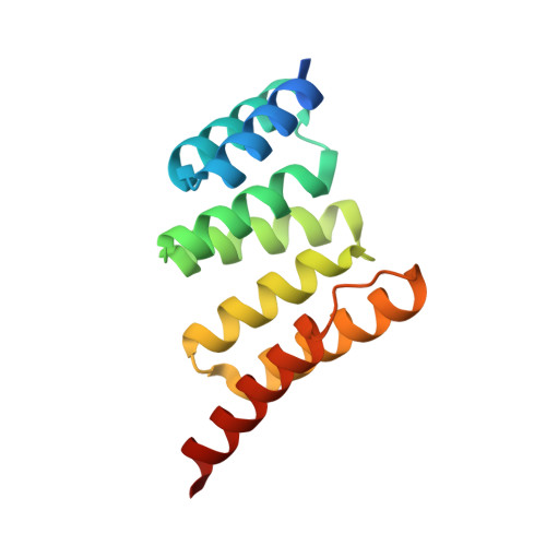
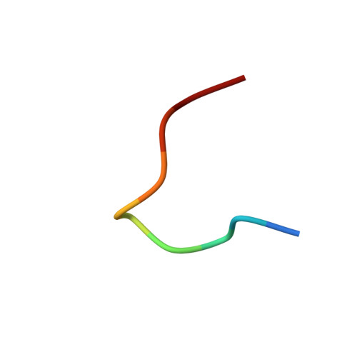
Discovery and Structure-Based Design of Macrocyclic Peptides Targeting STUB1.

Ng, S.Brueckner, A.C.Bahmanjah, S.Deng, Q.Johnston, J.M.Ge, L.Duggal, R.Habulihaz, B.Barlock, B.Ha, S.Sadruddin, A.Yeo, C.Strickland, C.Peier, A.Henry, B.Sherer, E.C.Partridge, A.W.

(2022) J Med Chem 

  • DOI: https://doi.org/10.1021/acs.jmedchem.2c00406
  • Primary Citation Related Structures: 
    7TB1

  • PubMed Abstract: 

    Recent evidence suggests that deletion of STUB1─a pivotal negative regulator of interferon-γ sensing─may potentially clear malignant cells. However, current studies rely primarily on genetic approaches, as pharmacological inhibitors of STUB1 are lacking. Identifying a tool compound will be a step toward validating the target in a broader therapeutic sense. Herein, screening more than a billion macrocyclic peptides resulted in STUB1 binders, which were further optimized by a structure-enabled in silico design. The strategy to replace the macrocyclic peptides' hydrophilic and solvent-exposed region with a hydrophobic scaffold improved cellular permeability while maintaining the binding conformation. Further substitution of the permeability-limiting terminal aspartic acid with a tetrazole bioisostere retained the binding to a certain extent while improving permeability, suggesting a path forward. Although not optimal for cellular study, the current lead provides a valuable template for further development into selective tool compounds for STUB1 to enable target validation.


  • Organizational Affiliation
    • Quantitative Biosciences, MSD, 8 Biomedical Grove, Singapore 138665.

Macromolecule Content 

  • Total Structure Weight: 33.91 kDa 
  • Atom Count: 2,468 
  • Modeled Residue Count: 273 
  • Deposited Residue Count: 300 
  • Unique protein chains: 2

Macromolecules

Find similar proteins by:|  3D Structure
Entity ID: 1
MoleculeChains  Sequence LengthOrganismDetailsImage
E3 ubiquitin-protein ligase CHIP
A, B
140Homo sapiensMutation(s): 0 
Gene Names: STUB1CHIPPP1131
EC: 2.3.2.27
UniProt & NIH Common Fund Data Resources
Find proteins for Q9UNE7 (Homo sapiens)
Explore Q9UNE7 
Go to UniProtKB:  Q9UNE7
PHAROS:  Q9UNE7
GTEx:  ENSG00000103266 
Entity Groups
Sequence Clusters30% Identity50% Identity70% Identity90% Identity95% Identity100% Identity
UniProt GroupQ9UNE7
Sequence Annotations
Expand
Reference Sequence
Find similar proteins by:  Sequence   |   3D Structure  
Entity ID: 2
MoleculeChains  Sequence LengthOrganismDetailsImage
ALA-CYS-SER-SER-ILE-TRP-CYS-PRO-ASP-GLY
C, D
10synthetic constructMutation(s): 0 

Experimental Data & Validation

Experimental Data

  • Method: X-RAY DIFFRACTION
  • Resolution: 1.78 Å
  • R-Value Free:  0.278 (Depositor), 0.267 (DCC) 
  • R-Value Work:  0.224 (Depositor), 0.215 (DCC) 
  • R-Value Observed: 0.227 (Depositor) 
Space Group: P 1 21 1
Unit Cell:
Length ( Å )Angle ( ˚ )
a = 36.011α = 90
b = 77.602β = 92.42
c = 46.354γ = 90
Software Package:
Software NamePurpose
BUSTERrefinement
PDB_EXTRACTdata extraction
XDSdata reduction
Aimlessdata scaling
PHASERphasing

Structure Validation

View Full Validation Report


Ligand Structure Quality Assessment 


Entry History 

& Funding Information

Deposition Data


Funding OrganizationLocationGrant Number
Not funded--

Revision History  (Full details and data files)

  • Version 1.0: 2022-07-27
    Type: Initial release
  • Version 1.1: 2023-10-18
    Changes: Data collection, Refinement description
  • Version 1.2: 2024-11-06
    Changes: Structure summary