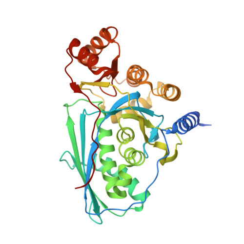
Crystal structure of mevalonate 3,5-bisphosphate decarboxylase reveals insight into the evolution of decarboxylases in the mevalonate metabolic pathways.
Aoki, M., Vinokur, J., Motoyama, K., Ishikawa, R., Collazo, M., Cascio, D., Sawaya, M.R., Ito, T., Bowie, J.U., Hemmi, H.(2022) J Biological Chem 298: 102111-102111
- PubMed: 35690147 Search on PubMedSearch on PubMed Central
- DOI: https://doi.org/10.1016/j.jbc.2022.102111
- Primary Citation Related Structures:
7T71 - PubMed Abstract:
Mevalonate 3,5-bisphosphate decarboxylase is involved in the recently discovered Thermoplasma-type mevalonate pathway. The enzyme catalyzes the elimination of the 3-phosphate group from mevalonate 3,5-bisphosphate as well as concomitant decarboxylation of the substrate. This entire reaction of the enzyme resembles the latter half-reactions of its homologs, diphosphomevalonate decarboxylase and phosphomevalonate decarboxylase, which also catalyze ATP-dependent phosphorylation of the 3-hydroxyl group of their substrates. However, the crystal structure of mevalonate 3,5-bisphosphate decarboxylase and the structural reasons of the difference between reactions catalyzed by the enzyme and its homologs are unknown. In this study, we determined the X-ray crystal structure of mevalonate 3,5-bisphosphate decarboxylase from Picrophilus torridus, a thermoacidophilic archaeon of the order Thermoplasmatales. Structural and mutational analysis demonstrated the importance of a conserved aspartate residue for enzyme activity. In addition, although crystallization was performed in the absence of substrate or ligands, residual electron density having the shape of a fatty acid was observed at a position overlapping the ATP-binding site of the homologous enzyme, diphosphomevalonate decarboxylase. This finding is in agreement with the expected evolutionary route from phosphomevalonate decarboxylase (ATP-dependent) to mevalonate 3,5-bisphosphate decarboxylase (ATP-independent) through the loss of kinase activity. We found that the binding of geranylgeranyl diphosphate, an intermediate of the archeal isoprenoid biosynthesis pathway, evoked significant activation of mevalonate 3,5-bisphosphate decarboxylase, and several mutations at the putative geranylgeranyl diphosphate-binding site impaired this activation, suggesting the physiological importance of ligand binding as well as a possible novel regulatory system employed by the Thermoplasma-type mevalonate pathway.
- Department of Applied Biosciences, Graduate School of Bioagricultural Sciences, Nagoya University, Furo-cho, Nagoya, Aichi, Japan.
Organizational Affiliation: