7T6W | pdb_00007t6w

Crystal structure of Chaetomium Glucosidase I (apo)


Experimental Data Snapshot

  • Method: X-RAY DIFFRACTION
  • Resolution: 2.60 Å
  • R-Value Free: 
    0.250 (Depositor), 0.250 (DCC) 
  • R-Value Work: 
    0.190 (Depositor), 0.190 (DCC) 
  • R-Value Observed: 
    0.193 (Depositor) 

Starting Model: experimental
View more details

wwPDB Validation 3D Report Full Report

Validation slider image for 7T6W

This is version 1.3 of the entry. See complete history

Literature

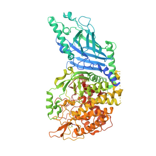
Identification of Endoplasmic Reticulum alpha-Glucosidase I from a Thermophilic Fungus as a Platform for Structure-Guided Antiviral Drug Design.

Karade, S.S.Kolesnikov, A.Treston, A.M.Mariuzza, R.A.

(2022) Biochemistry 61: 822-832

  • DOI: https://doi.org/10.1021/acs.biochem.2c00092
  • Primary Citation Related Structures: 
    7T66, 7T68, 7T6W, 7T8V

  • PubMed Abstract: 

    All viruses depend on host cell proteins for replication. Denying viruses' access to the function of critical host proteins can result in antiviral activity against multiple virus families. In particular, small-molecule drug candidates which inhibit the α-glucosidase enzymes of the endoplasmic reticulum (ER) translation quality control (QC) pathway have demonstrated broad-spectrum antiviral activities and low risk for development of viral resistance. However, antiviral drug discovery focused on the ERQC enzyme α-glucosidase I (α-GluI) has been hampered by difficulties in obtaining crystal structures of complexes with inhibitors. We report here the identification of an orthologous enzyme from a thermophilic fungus, Chaetomium thermophilum ( Ct ), as a robust surrogate for mammalian ER α-GluI and a platform for inhibitor design. Previously annotated only as a hypothetical protein, the Ct protein was validated as a bona fide α-glucosidase by comparing its crystal structure to that of mammalian α-GluI, by demonstrating enzymatic activity on the unusual α-d-Glc p -(1 → 2)-α-d-Glc p -(1 → 3) substrate glycan, and by showing that well-known inhibitors of mammalian α-GluI (1-DNJ, UV-4, UV-5) also inhibit Ct α-GluI. Crystal structures of Ct α-GluI in complex with three such inhibitors (UV-4, UV-5, EB-0159) revealed extensive interactions with all four enzyme subsites and provided insights into the catalytic mechanism. Identification of ER Ct α-GluI as a surrogate for mammalian α-GluI will accelerate the structure-guided discovery of broad-spectrum antivirals. This study also highlights Ct as a source of thermostable eukaryotic proteins, such as ER α-Glu I, that lack orthologs in bacterial or archaeal thermophiles.


  • Organizational Affiliation
    • Institute for Bioscience and Biotechnology Research, University of Maryland, Rockville, Maryland 20850, United States.

Macromolecule Content 

  • Total Structure Weight: 188.48 kDa 
  • Atom Count: 12,174 
  • Modeled Residue Count: 1,532 
  • Deposited Residue Count: 1,638 
  • Unique protein chains: 1

Macromolecules

Find similar proteins by:|  3D Structure
Entity ID: 1
MoleculeChains  Sequence LengthOrganismDetailsImage
Chaetomium alpha glucosidase
A, B
819Thermochaetoides thermophila DSM 1495Mutation(s): 0 
Gene Names: CTHT_0061620
EC: 3.2.1.106
UniProt
Find proteins for G0SFD1 (Chaetomium thermophilum (strain DSM 1495 / CBS 144.50 / IMI 039719))
Explore G0SFD1 
Go to UniProtKB:  G0SFD1
Entity Groups
Sequence Clusters30% Identity50% Identity70% Identity90% Identity95% Identity100% Identity
UniProt GroupG0SFD1
Glycosylation
Glycosylation Sites: 1
Sequence Annotations
Expand
Reference Sequence

Oligosaccharides

Help  
Entity ID: 2
MoleculeChains Length2D Diagram GlycosylationD Interactions
2-acetamido-2-deoxy-beta-D-glucopyranose-(1-4)-2-acetamido-2-deoxy-beta-D-glucopyranose
C
2N-Glycosylation
Glycosylation Resources
GlyTouCan: G42666HT
GlyCosmos: G42666HT
GlyGen: G42666HT

Small Molecules

Ligands 2 Unique
IDChains Name / Formula / InChI Key2D Diagram3D Interactions
SO4

Query on SO4


Download:Ideal Coordinates CCD File
G [auth A]
H [auth A]
I [auth A]
J [auth A]
K [auth A]
G [auth A],
H [auth A],
I [auth A],
J [auth A],
K [auth A],
L [auth A],
P [auth B],
Q [auth B],
R [auth B]
SULFATE ION
O4 S
QAOWNCQODCNURD-UHFFFAOYSA-L
GOL

Query on GOL


Download:Ideal Coordinates CCD File
D [auth A]
E [auth A]
F [auth A]
M [auth B]
N [auth B]
D [auth A],
E [auth A],
F [auth A],
M [auth B],
N [auth B],
O [auth B]
GLYCEROL
C3 H8 O3
PEDCQBHIVMGVHV-UHFFFAOYSA-N

Experimental Data & Validation

Experimental Data

  • Method: X-RAY DIFFRACTION
  • Resolution: 2.60 Å
  • R-Value Free:  0.250 (Depositor), 0.250 (DCC) 
  • R-Value Work:  0.190 (Depositor), 0.190 (DCC) 
  • R-Value Observed: 0.193 (Depositor) 
Space Group: I 2 2 2
Unit Cell:
Length ( Å )Angle ( ˚ )
a = 139.152α = 90
b = 178.804β = 90
c = 180.697γ = 90
Software Package:
Software NamePurpose
REFMACrefinement
PDB_EXTRACTdata extraction
HKL-2000data reduction
HKL-2000data scaling
PHASERphasing

Structure Validation

View Full Validation Report


Entry History 

& Funding Information

Deposition Data


Funding OrganizationLocationGrant Number
Not funded--

Revision History  (Full details and data files)

  • Version 1.0: 2022-05-04
    Type: Initial release
  • Version 1.1: 2022-05-25
    Changes: Database references
  • Version 1.2: 2023-10-18
    Changes: Data collection, Refinement description
  • Version 1.3: 2024-11-06
    Changes: Structure summary