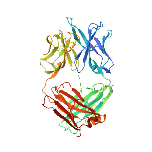
7T62 | pdb_00007t62

GPC2 HEP CT3 complex


Experimental Data Snapshot

  • Method: ELECTRON MICROSCOPY
  • Resolution: 21.0 Å
  • Aggregation State: PARTICLE 
  • Reconstruction Method: SINGLE PARTICLE 

wwPDB Validation 3D Report Full Report

Validation slider image for 7T62

This is version 1.1 of the entry. See complete history. 

Literature

CAR T cells targeting tumor-associated exons of glypican 2 regress neuroblastoma in mice.

Li, N., Torres, M.B., Spetz, M.R., Wang, R., Peng, L., Tian, M., Dower, C.M., Nguyen, R., Sun, M., Tai, C.H., de Val, N., Cachau, R., Wu, X., Hewitt, S.M., Kaplan, R.N., Khan, J., St Croix, B., Thiele, C.J., Ho, M.

(2021) Cell Rep Med 2: 100297-100297

  • DOI: https://doi.org/10.1016/j.xcrm.2021.100297
  • Primary Citation Related Structures: 
    7T62

  • PubMed Abstract: 

    Targeting solid tumors must overcome several major obstacles, in particular, the identification of elusive tumor-specific antigens. Here, we devise a strategy to help identify tumor-specific epitopes. Glypican 2 (GPC2) is overexpressed in neuroblastoma. Using RNA sequencing (RNA-seq) analysis, we show that exon 3 and exons 7-10 of GPC2 are expressed in cancer but are minimally expressed in normal tissues. Accordingly, we discover a monoclonal antibody (CT3) that binds exons 3 and 10 and visualize the complex structure of CT3 and GPC2 by electron microscopy. The potential of this approach is exemplified by designing CT3-derived chimeric antigen receptor (CAR) T cells that regress neuroblastoma in mice. Genomic sequencing of T cells recovered from mice reveals the CAR integration sites that may contribute to CAR T cell proliferation and persistence. These studies demonstrate how RNA-seq data can be exploited to help identify tumor-associated exons that can be targeted by CAR T cell therapies.


  • Organizational Affiliation: 
    • Laboratory of Molecular Biology, Center for Cancer Research, National Cancer Institute, National Institutes of Health, Bethesda, MD 20892, USA.

Macromolecule Content 

  • Total Structure Weight: 108.36 kDa 
  • Atom Count: 7,603 
  • Modeled Residue Count: 993 
  • Deposited Residue Count: 994 
  • Unique protein chains: 2

Macromolecules

Find similar proteins by:|  3D Structure
Entity ID: 1
MoleculeChains  Sequence LengthOrganismDetailsImage
Glypican-2561Homo sapiensMutation(s): 0 
Gene Names: GPC2
UniProt & NIH Common Fund Data Resources
Find proteins for Q8N158 (Homo sapiens)
Explore Q8N158 
Go to UniProtKB:  Q8N158
PHAROS:  Q8N158
GTEx:  ENSG00000213420 
Entity Groups
Sequence Clusters30% Identity50% Identity70% Identity90% Identity95% Identity100% Identity
UniProt GroupQ8N158
Sequence Annotations
Expand
Reference Sequence
Find similar proteins by:|  3D Structure
Entity ID: 2
MoleculeChains  Sequence LengthOrganismDetailsImage
CT3433Mus musculusMutation(s): 0 
Entity Groups
Sequence Clusters30% Identity50% Identity70% Identity90% Identity95% Identity100% Identity
Sequence Annotations
Expand
Reference Sequence

Experimental Data & Validation

Experimental Data

  • Method: ELECTRON MICROSCOPY
  • Resolution: 21.0 Å
  • Aggregation State: PARTICLE 
  • Reconstruction Method: SINGLE PARTICLE 
EM Software:
TaskSoftware PackageVersion
RECONSTRUCTIONRELION3.08

Structure Validation

View Full Validation Report


Entry History 

& Funding Information

Deposition Data


Funding OrganizationLocationGrant Number
National Institutes of Health/National Cancer Institute (NIH/NCI)United StatesZ01 BC010891
National Institutes of Health/National Cancer Institute (NIH/NCI)United StatesCancer Moonshot Program

Revision History  (Full details and data files)

  • Version 1.0: 2021-12-22
    Type: Initial release
  • Version 1.1: 2024-10-23
    Changes: Data collection, Database references, Structure summary