


Structure of bacteriophage lambda tube protein V in C6
Prokhorov, N.S., Yang, Q., Catalano, C.E., Morais, M.C.To be published.
Experimental Data Snapshot
wwPDB Validation 3D Report Full Report

Entity ID: 1 | |||||
|---|---|---|---|---|---|
| Molecule | Chains | Sequence Length | Organism | Details | Image |
| Tail tube protein | 245 | Lambdavirus lambda | Mutation(s): 0 Gene Names: V, lambdap13 | 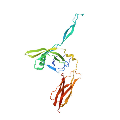 | |
UniProt | |||||
Entity Groups | |||||
| Sequence Clusters | 30% Identity50% Identity70% Identity90% Identity95% Identity100% Identity | ||||
| UniProt Group | P03733 | ||||
Sequence AnnotationsExpand | |||||
Reference Sequence | |||||
| Funding Organization | Location | Grant Number |
|---|---|---|
| National Institutes of Health/National Institute of General Medical Sciences (NIH/NIGMS) | United States | R01GM127365 |


