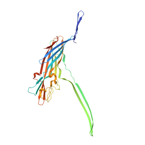
Enterococci are a part of human microbiota and a leading cause of multidrug resistant infections. Here, we identify a family of Enterococcus pore-forming toxins (Epxs) in E. faecalis, E. faecium, and E. hirae strains isolated across the globe. Structural studies reveal that Epxs form a branch of β-barrel pore-forming toxins with a β-barrel protrusion (designated the top domain) sitting atop the cap domain. Through a genome-wide CRISPR-Cas9 screen, we identify human leukocyte antigen class I (HLA-I) complex as a receptor for two members (Epx2 and Epx3), which preferentially recognize human HLA-I and homologous MHC-I of equine, bovine, and porcine, but not murine, origin. Interferon exposure, which stimulates MHC-I expression, sensitizes human cells and intestinal organoids to Epx2 and Epx3 toxicity. Co-culture with Epx2-harboring E. faecium damages human peripheral blood mononuclear cells and intestinal organoids, and this toxicity is neutralized by an Epx2 antibody, demonstrating the toxin-mediated virulence of Epx-carrying Enterococcus.
Organizational Affiliation:
Department of Urology, Boston Children's Hospital, Department of Surgery, Harvard Medical School, Boston, MA 02115, USA; Department of Microbiology, Harvard Medical School, Boston, MA 02115, USA.
Department of Microbiology, Harvard Medical School, Boston, MA 02115, USA.
Department of Microbiology, Harvard Medical School, Boston, MA 02115, USA; Department of Ophthalmology, Harvard Medical School, Massachusetts Eye and Ear Infirmary, Boston, MA 02114, USA; Broad Institute of MIT and Harvard, Cambridge, MA 02142, USA; Multidrug-Resistant Organism Repository and Surveillance Network (MRSN), Walter Reed Army Institute of Research, Silver Spring, MD 20910, USA.
Department of Molecular Medicine, The Scripps Research Institute, Jupiter, FL 33458, USA.
Wyss Institute for Biologically Inspired Engineering, Harvard University, Boston, MA 02115.
Department of Urology, Boston Children's Hospital, Department of Surgery, Harvard Medical School, Boston, MA 02115, USA.
Division of Endocrinology, Boston Children's Hospital, and Department of Pediatrics, Harvard Medical School, Boston, MA 02115, USA.
Program in Cellular and Molecular Medicine, Boston Children's Hospital, and Department of Biological Chemistry and Molecular Pharmacology, Harvard Medical School, Boston, MA 02115, USA; Department of Immunology, University of Connecticut Health School of Medicine, Farmington, CT 06030, USA.
Vascular Biology Program, Department of Surgery, Boston Children's Hospital and Harvard Medical School, Boston, MA 02115, USA.
Division of Endocrinology, Boston Children's Hospital, and Department of Pediatrics, Harvard Medical School, Boston, MA 02115, USA; Harvard Stem Cell Institute, Cambridge, MA 02138, USA.
Program in Cellular and Molecular Medicine, Boston Children's Hospital, and Department of Biological Chemistry and Molecular Pharmacology, Harvard Medical School, Boston, MA 02115, USA.
Broad Institute of MIT and Harvard, Cambridge, MA 02142, USA.
Department of Microbiology, Harvard Medical School, Boston, MA 02115, USA; Department of Ophthalmology, Harvard Medical School, Massachusetts Eye and Ear Infirmary, Boston, MA 02114, USA; Broad Institute of MIT and Harvard, Cambridge, MA 02142, USA. Electronic address: michael_gilmore@meei.harvard.edu.
Department of Microbiology, Harvard Medical School, Boston, MA 02115, USA; Broad Institute of MIT and Harvard, Cambridge, MA 02142, USA; Division of Infectious Diseases, Department of Medicine, Brigham and Women's Hospital, Boston, MA 02115, USA. Electronic address: jonathan_abraham@hms.harvard.edu.
Department of Urology, Boston Children's Hospital, Department of Surgery, Harvard Medical School, Boston, MA 02115, USA; Department of Microbiology, Harvard Medical School, Boston, MA 02115, USA. Electronic address: min.dong@childrens.harvard.edu.