7T2T | pdb_00007t2t

SARS-CoV2 Mpro native form


Experimental Data Snapshot

  • Method: X-RAY DIFFRACTION
  • Resolution: 1.45 Å
  • R-Value Free: 
    0.200 (Depositor), 0.206 (DCC) 
  • R-Value Work: 
    0.155 (Depositor), 0.161 (DCC) 
  • R-Value Observed: 
    0.157 (Depositor) 

Starting Model: experimental
View more details

wwPDB Validation 3D Report Full Report

Validation slider image for 7T2T

This is version 1.2 of the entry. See complete history

Literature

Structural and functional characterization of NEMO cleavage by SARS-CoV-2 3CLpro.

Hameedi, M.A.T Prates, E.Garvin, M.R.Mathews, I.I.Amos, B.K.Demerdash, O.Bechthold, M.Iyer, M.Rahighi, S.Kneller, D.W.Kovalevsky, A.Irle, S.Vuong, V.Q.Mitchell, J.C.Labbe, A.Galanie, S.Wakatsuki, S.Jacobson, D.

(2022) Nat Commun 13: 5285-5285

  • DOI: https://doi.org/10.1038/s41467-022-32922-9
  • Primary Citation Related Structures: 
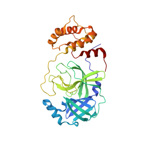
    7T2T, 7T2U, 7T2V

  • PubMed Abstract: 

    In addition to its essential role in viral polyprotein processing, the SARS-CoV-2 3C-like protease (3CLpro) can cleave human immune signaling proteins, like NF-κB Essential Modulator (NEMO) and deregulate the host immune response. Here, in vitro assays show that SARS-CoV-2 3CLpro cleaves NEMO with fine-tuned efficiency. Analysis of the 2.50 Å resolution crystal structure of 3CLpro C145S bound to NEMO 226-234 reveals subsites that tolerate a range of viral and host substrates through main chain hydrogen bonds while also enforcing specificity using side chain hydrogen bonds and hydrophobic contacts. Machine learning- and physics-based computational methods predict that variation in key binding residues of 3CLpro-NEMO helps explain the high fitness of SARS-CoV-2 in humans. We posit that cleavage of NEMO is an important piece of information to be accounted for, in the pathology of COVID-19.


  • Organizational Affiliation
    • SLAC National Accelerator Laboratory, Stanford Synchrotron Radiation Lightsource, Structural Molecular Biology, Menlo Park, CA, 94025, USA.

Macromolecule Content 

  • Total Structure Weight: 33.83 kDa 
  • Atom Count: 2,630 
  • Modeled Residue Count: 305 
  • Deposited Residue Count: 306 
  • Unique protein chains: 1

Macromolecules

Find similar proteins by:|  3D Structure
Entity ID: 1
MoleculeChains  Sequence LengthOrganismDetailsImage
3C-like proteinase306Severe acute respiratory syndrome coronavirus 2Mutation(s): 0 
Gene Names: rep1a-1b
EC: 3.4.22.69
UniProt
Find proteins for P0DTD1 (Severe acute respiratory syndrome coronavirus 2)
Explore P0DTD1 
Go to UniProtKB:  P0DTD1
Entity Groups
Sequence Clusters30% Identity50% Identity70% Identity90% Identity95% Identity100% Identity
UniProt GroupP0DTD1
Sequence Annotations
Expand
Reference Sequence

Experimental Data & Validation

Experimental Data

  • Method: X-RAY DIFFRACTION
  • Resolution: 1.45 Å
  • R-Value Free:  0.200 (Depositor), 0.206 (DCC) 
  • R-Value Work:  0.155 (Depositor), 0.161 (DCC) 
  • R-Value Observed: 0.157 (Depositor) 
Space Group: C 1 2 1
Unit Cell:
Length ( Å )Angle ( ˚ )
a = 114.17α = 90
b = 53.44β = 103.02
c = 44.87γ = 90
Software Package:
Software NamePurpose
XSCALEdata scaling
REFMACrefinement
PDB_EXTRACTdata extraction
XDSdata reduction
MOLREPphasing

Structure Validation

View Full Validation Report


Entry History 

& Funding Information

Deposition Data


Funding OrganizationLocationGrant Number
Department of Energy (DOE, United States)United StatesDE-AC02-76SF00515

Revision History  (Full details and data files)

  • Version 1.0: 2022-09-14
    Type: Initial release
  • Version 1.1: 2022-09-21
    Changes: Database references
  • Version 1.2: 2023-10-18
    Changes: Data collection, Refinement description