Novel sterol binding domains in bacteria.
Zhai, L., Bonds, A.C., Smith, C.A., Oo, H., Chou, J.C., Welander, P.V., Dassama, L.M.K.(2024) Elife 12
- PubMed: 38329015 Search on PubMedSearch on PubMed Central
- DOI: https://doi.org/10.7554/eLife.90696
- Primary Citation Related Structures:
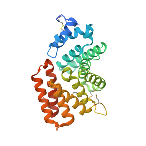
7T1M, 7T1S - PubMed Abstract:
Sterol lipids are widely present in eukaryotes and play essential roles in signaling and modulating membrane fluidity. Although rare, some bacteria also produce sterols, but their function in bacteria is not known. Moreover, many more species, including pathogens and commensal microbes, acquire or modify sterols from eukaryotic hosts through poorly understood molecular mechanisms. The aerobic methanotroph Methylococcus capsulatus was the first bacterium shown to synthesize sterols, producing a mixture of C-4 methylated sterols that are distinct from those observed in eukaryotes. C-4 methylated sterols are synthesized in the cytosol and localized to the outer membrane, suggesting that a bacterial sterol transport machinery exists. Until now, the identity of such machinery remained a mystery. In this study, we identified three novel proteins that may be the first examples of transporters for bacterial sterol lipids. The proteins, which all belong to well-studied families of bacterial metabolite transporters, are predicted to reside in the inner membrane, periplasm, and outer membrane of M. capsulatus, and may work as a conduit to move modified sterols to the outer membrane. Quantitative analysis of ligand binding revealed their remarkable specificity for 4-methylsterols, and crystallographic structures coupled with docking and molecular dynamics simulations revealed the structural bases for substrate binding by two of the putative transporters. Their striking structural divergence from eukaryotic sterol transporters signals that they form a distinct sterol transport system within the bacterial domain. Finally, bioinformatics revealed the widespread presence of similar transporters in bacterial genomes, including in some pathogens that use host sterol lipids to construct their cell envelopes. The unique folds of these bacterial sterol binding proteins should now guide the discovery of other proteins that handle this essential metabolite.
- Department of Chemistry and Sarafan ChEM-H, Stanford University, Stanford, United States.
Organizational Affiliation: