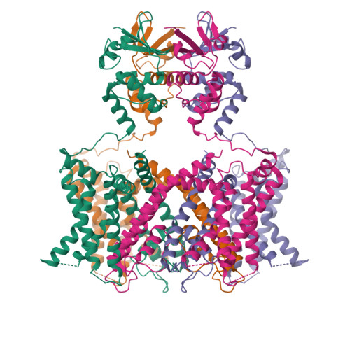
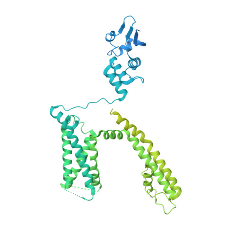
7SSY

Structure of human Kv1.3 (alternate conformation)


Experimental Data Snapshot

  • Method: ELECTRON MICROSCOPY
  • Resolution: 2.89 Å
  • Aggregation State: PARTICLE 
  • Reconstruction Method: SINGLE PARTICLE 

wwPDB Validation   3D Report Full Report


This is version 1.2 of the entry. See complete history


Literature

Structures of the T cell potassium channel Kv1.3 with immunoglobulin modulators.

Selvakumar, P.Fernandez-Marino, A.I.Khanra, N.He, C.Paquette, A.J.Wang, B.Huang, R.Smider, V.V.Rice, W.J.Swartz, K.J.Meyerson, J.R.

(2022) Nat Commun 13: 3854-3854

  • DOI: https://doi.org/10.1038/s41467-022-31285-5
  • Primary Citation of Related Structures:  
    7SSV, 7SSX, 7SSY, 7SSZ, 8DFL

  • PubMed Abstract: 

    The Kv1.3 potassium channel is expressed abundantly on activated T cells and mediates the cellular immune response. This role has made the channel a target for therapeutic immunomodulation to block its activity and suppress T cell activation. Here, we report structures of human Kv1.3 alone, with a nanobody inhibitor, and with an antibody-toxin fusion blocker. Rather than block the channel directly, four copies of the nanobody bind the tetramer's voltage sensing domains and the pore domain to induce an inactive pore conformation. In contrast, the antibody-toxin fusion docks its toxin domain at the extracellular mouth of the channel to insert a critical lysine into the pore. The lysine stabilizes an active conformation of the pore yet blocks ion permeation. This study visualizes Kv1.3 pore dynamics, defines two distinct mechanisms to suppress Kv1.3 channel activity with exogenous inhibitors, and provides a framework to aid development of emerging T cell immunotherapies.


  • Organizational Affiliation

    Department of Physiology and Biophysics, Weill Cornell Medical College, New York, NY, USA.


Macromolecules
Find similar proteins by:  (by identity cutoff)  |  3D Structure
Entity ID: 1
MoleculeChains Sequence LengthOrganismDetailsImage
Potassium voltage-gated channel subfamily A member 3,Green fluorescent protein fusion
A, B, C, D
856Homo sapiensAequorea victoria
This entity is chimeric
Mutation(s): 0 
Gene Names: KCNA3HGK5GFP
Membrane Entity: Yes 
UniProt & NIH Common Fund Data Resources
Find proteins for P42212 (Aequorea victoria)
Explore P42212 
Go to UniProtKB:  P42212
Find proteins for P22001 (Homo sapiens)
Explore P22001 
Go to UniProtKB:  P22001
PHAROS:  P22001
GTEx:  ENSG00000177272 
Entity Groups  
Sequence Clusters30% Identity50% Identity70% Identity90% Identity95% Identity100% Identity
UniProt GroupsP22001P42212
Sequence Annotations
Expand
  • Reference Sequence
Experimental Data & Validation

Experimental Data

  • Method: ELECTRON MICROSCOPY
  • Resolution: 2.89 Å
  • Aggregation State: PARTICLE 
  • Reconstruction Method: SINGLE PARTICLE 

Structure Validation

View Full Validation Report


Entry History & Funding Information

Deposition Data


Funding OrganizationLocationGrant Number
Not fundedUnited States--

Revision History  (Full details and data files)

  • Version 1.0: 2022-06-29
    Type: Initial release
  • Version 1.1: 2022-07-20
    Changes: Database references
  • Version 1.2: 2024-06-05
    Changes: Data collection