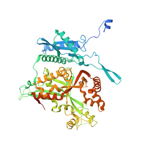
7SQR | pdb_00007sqr

201phi2-1 Chimallin localized tetramer reconstruction


Experimental Data Snapshot

  • Method: ELECTRON MICROSCOPY
  • Resolution: 3.40 Å
  • Aggregation State: PARTICLE 
  • Reconstruction Method: SINGLE PARTICLE 

wwPDB Validation 3D Report Full Report

Validation slider image for 7SQR

This is version 1.3 of the entry. See complete history. 

Literature

Architecture and self-assembly of the jumbo bacteriophage nuclear shell.

Laughlin, T.G., Deep, A., Prichard, A.M., Seitz, C., Gu, Y., Enustun, E., Suslov, S., Khanna, K., Birkholz, E.A., Armbruster, E., McCammon, J.A., Amaro, R.E., Pogliano, J., Corbett, K.D., Villa, E.

(2022) Nature 608: 429-435

  • DOI: https://doi.org/10.1038/s41586-022-05013-4
  • Primary Citation Related Structures: 
    7SQQ, 7SQR, 7SQS, 7SQT, 7SQU, 7SQV

  • PubMed Abstract: 

    Bacteria encode myriad defences that target the genomes of infecting bacteriophage, including restriction-modification and CRISPR-Cas systems 1 . In response, one family of large bacteriophages uses a nucleus-like compartment to protect its replicating genomes by excluding host defence factors 2-4 . However, the principal composition and structure of this compartment remain unknown. Here we find that the bacteriophage nuclear shell assembles primarily from one protein, which we name chimallin (ChmA). Combining cryo-electron tomography of nuclear shells in bacteriophage-infected cells and cryo-electron microscopy of a minimal chimallin compartment in vitro, we show that chimallin self-assembles as a flexible sheet into closed micrometre-scale compartments. The architecture and assembly dynamics of the chimallin shell suggest mechanisms for its nucleation and growth, and its role as a scaffold for phage-encoded factors mediating macromolecular transport, cytoskeletal interactions, and viral maturation.


  • Organizational Affiliation: 
    • Department of Molecular Biology, School of Biological Sciences, University of California San Diego, La Jolla, CA, USA.

Macromolecule Content 

  • Total Structure Weight: 837.41 kDa 
  • Atom Count: 17,272 
  • Modeled Residue Count: 2,216 
  • Deposited Residue Count: 7,608 
  • Unique protein chains: 1

Macromolecules

Find similar proteins by:|  3D Structure
Entity ID: 1
MoleculeChains  Sequence LengthOrganismDetailsImage
Chimallin
A, B, C, D, E
A, B, C, D, E, F, G, H, I, J, K, L
634Pseudomonas phage 201phi2-1Mutation(s): 0 
Gene Names: 201phi2-1p105
UniProt
Find proteins for B3FIW8 (Pseudomonas phage 201phi2-1)
Explore B3FIW8 
Go to UniProtKB:  B3FIW8
Entity Groups
Sequence Clusters30% Identity50% Identity70% Identity90% Identity95% Identity100% Identity
UniProt GroupB3FIW8
Sequence Annotations
Expand
Reference Sequence

Experimental Data & Validation

Experimental Data

  • Method: ELECTRON MICROSCOPY
  • Resolution: 3.40 Å
  • Aggregation State: PARTICLE 
  • Reconstruction Method: SINGLE PARTICLE 
EM Software:
TaskSoftware PackageVersion
RECONSTRUCTIONRELION
MODEL REFINEMENTPHENIX

Structure Validation

View Full Validation Report


Entry History 

& Funding Information

Deposition Data


Funding OrganizationLocationGrant Number
National Institutes of Health/National Institute of General Medical Sciences (NIH/NIGMS)United StatesR01GM129245
National Institutes of Health/National Institute Of Allergy and Infectious Diseases (NIH/NIAID)United StatesR21AI148814
National Science Foundation (NSF, United States)United StatesNSF DBI 1920374
Howard Hughes Medical Institute (HHMI)United States--

Revision History  (Full details and data files)

  • Version 1.0: 2022-07-27
    Type: Initial release
  • Version 1.1: 2022-08-17
    Changes: Database references
  • Version 1.2: 2022-08-24
    Changes: Database references
  • Version 1.3: 2024-06-05
    Changes: Data collection