Flagellin outer domain dimerization modulates motility in pathogenic and soil bacteria from viscous environments.
Kreutzberger, M.A.B., Sobe, R.C., Sauder, A.B., Chatterjee, S., Pena, A., Wang, F., Giron, J.A., Kiessling, V., Costa, T.R.D., Conticello, V.P., Frankel, G., Kendall, M.M., Scharf, B.E., Egelman, E.H.(2022) Nat Commun 13: 1422-1422
- PubMed: 35301306 Search on PubMedSearch on PubMed Central
- DOI: https://doi.org/10.1038/s41467-022-29069-y
- Primary Citation Related Structures:
7SN4, 7SN7, 7SN9, 7SQD, 7SQJ - PubMed Abstract:
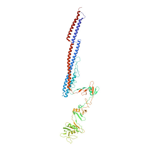
Flagellar filaments function as the propellers of the bacterial flagellum and their supercoiling is key to motility. The outer domains on the surface of the filament are non-critical for motility in many bacteria and their structures and functions are not conserved. Here, we show the atomic cryo-electron microscopy structures for flagellar filaments from enterohemorrhagic Escherichia coli O157:H7, enteropathogenic E. coli O127:H6, Achromobacter, and Sinorhizobium meliloti, where the outer domains dimerize or tetramerize to form either a sheath or a screw-like surface. These dimers are formed by 180° rotations of half of the outer domains. The outer domain sheath (ODS) plays a role in bacterial motility by stabilizing an intermediate waveform and prolonging the tumbling of E. coli cells. Bacteria with these ODS and screw-like flagellar filaments are commonly found in soil and human intestinal environments of relatively high viscosity suggesting a role for the dimerization in these environments.
- Department of Biochemistry and Molecular Genetics, University of Virginia School of Medicine, Charlottesville, VA, 22903, USA.
Organizational Affiliation: