7SO0 | pdb_00007so0

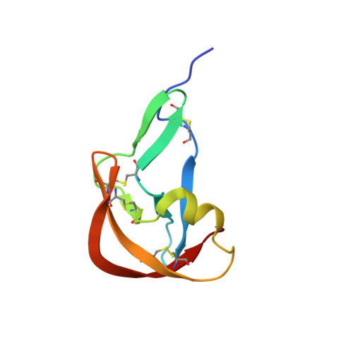
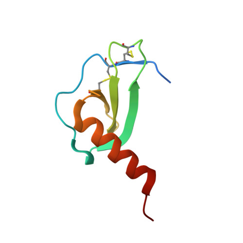
Crystal Structure of the Engineered Tick Evasin EVA-P974(F31A) Complexed to Human Chemokine CCL2


Experimental Data Snapshot

  • Method: X-RAY DIFFRACTION
  • Resolution: 1.74 Å
  • R-Value Free: 
    0.202 (Depositor), 0.203 (DCC) 
  • R-Value Work: 
    0.184 (Depositor), 0.185 (DCC) 
  • R-Value Observed: 
    0.185 (Depositor) 

Starting Model: experimental
View more details

wwPDB Validation 3D Report Full Report

Validation slider image for 7SO0

This is version 1.2 of the entry. See complete history. 

Literature

Structure-guided engineering of tick evasins for targeting chemokines in inflammatory diseases.

Bhusal, R.P., Aryal, P., Devkota, S.R., Pokhrel, R., Gunzburg, M.J., Foster, S.R., Lim, H.D., Payne, R.J., Wilce, M.C.J., Stone, M.J.

(2022) Proc Natl Acad Sci U S A 119

  • DOI: https://doi.org/10.1073/pnas.2122105119
  • Primary Citation Related Structures: 
    7S4N, 7S58, 7S59, 7S5A, 7SO0

  • PubMed Abstract: 

    As natural chemokine inhibitors, evasin proteins produced in tick saliva are potential therapeutic agents for numerous inflammatory diseases. Engineering evasins to block the desired chemokines and avoid off-target side effects requires structural understanding of their target selectivity. Structures of the class A evasin EVA-P974 bound to human CC chemokine ligands 7 and 17 (CCL7 and CCL17) and to a CCL8-CCL7 chimera reveal that the specificity of class A evasins for chemokines of the CC subfamily is defined by conserved, rigid backbone-backbone interactions, whereas the preference for a subset of CC chemokines is controlled by side-chain interactions at four hotspots in flexible structural elements. Hotspot mutations alter target preference, enabling inhibition of selected chemokines. The structure of an engineered EVA-P974 bound to CCL2 reveals an underlying molecular mechanism of EVA-P974 target preference. These results provide a structure-based framework for engineering evasins as targeted antiinflammatory therapeutics.


  • Organizational Affiliation: 
    • Monash Biomedicine Discovery Institute, Monash University, Clayton, VIC 3800, Australia; ram.bhusal@monash.edu martin.stone@monash.edu.

Macromolecule Content 

  • Total Structure Weight: 18.3 kDa 
  • Atom Count: 1,288 
  • Modeled Residue Count: 154 
  • Deposited Residue Count: 161 
  • Unique protein chains: 2

Macromolecules

Find similar proteins by:|  3D Structure
Entity ID: 1
MoleculeChains  Sequence LengthOrganismDetailsImage
Evasin P97485Amblyomma cajennenseMutation(s): 1 
UniProt
Find proteins for A0A023FDY8 (Amblyomma cajennense)
Explore A0A023FDY8 
Go to UniProtKB:  A0A023FDY8
Entity Groups
Sequence Clusters30% Identity50% Identity70% Identity90% Identity95% Identity100% Identity
UniProt GroupA0A023FDY8
Sequence Annotations
Expand
Reference Sequence
Find similar proteins by:|  3D Structure
Entity ID: 2
MoleculeChains  Sequence LengthOrganismDetailsImage
C-C motif chemokine 276Homo sapiensMutation(s): 0 
Gene Names: CCL2, MCP1, SCYA2
UniProt & NIH Common Fund Data Resources
Find proteins for P13500 (Homo sapiens)
Explore P13500 
Go to UniProtKB:  P13500
PHAROS:  P13500
GTEx:  ENSG00000108691 
Entity Groups
Sequence Clusters30% Identity50% Identity70% Identity90% Identity95% Identity100% Identity
UniProt GroupP13500
Sequence Annotations
Expand
Reference Sequence

Experimental Data & Validation

Experimental Data

  • Method: X-RAY DIFFRACTION
  • Resolution: 1.74 Å
  • R-Value Free:  0.202 (Depositor), 0.203 (DCC) 
  • R-Value Work:  0.184 (Depositor), 0.185 (DCC) 
  • R-Value Observed: 0.185 (Depositor) 
Space Group: P 32
Unit Cell:
Length ( Å )Angle ( ˚ )
a = 36.785α = 90
b = 36.785β = 90
c = 108.682γ = 120
Software Package:
Software NamePurpose
PHENIXrefinement
XDSdata reduction
Aimlessdata scaling
PHASERphasing

Structure Validation

View Full Validation Report


Entry History 

& Funding Information

Deposition Data


Funding OrganizationLocationGrant Number
National Health and Medical Research Council (NHMRC, Australia)Australia--

Revision History  (Full details and data files)

  • Version 1.0: 2022-03-16
    Type: Initial release
  • Version 1.1: 2023-10-18
    Changes: Data collection, Refinement description
  • Version 1.2: 2024-10-23
    Changes: Structure summary