7SKW | pdb_00007skw

Ab initio structure of triclinic lysozyme from electron-counted MicroED data


Experimental Data Snapshot

  • Method: ELECTRON CRYSTALLOGRAPHY
  • Resolution: 0.87 Å
  • R-Value Free: 
    0.221 (Depositor) 
  • R-Value Work: 
    0.197 (Depositor) 

wwPDB Validation 3D Report Full Report

Validation slider image for 7SKW

This is version 1.3 of the entry. See complete history

Literature

Ab initio phasing macromolecular structures using electron-counted MicroED data.

Martynowycz, M.W.Clabbers, M.T.B.Hattne, J.Gonen, T.

(2022) Nat Methods 19: 724-729

  • DOI: https://doi.org/10.1038/s41592-022-01485-4
  • Primary Citation Related Structures: 
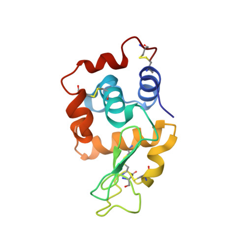
    7SKW, 7SKX

  • PubMed Abstract: 

    Structures of two globular proteins were determined ab initio using microcrystal electron diffraction (MicroED) data that were collected on a direct electron detector in counting mode. Microcrystals were identified using a scanning electron microscope (SEM) and thinned with a focused ion beam (FIB) to produce crystalline lamellae of ideal thickness. Continuous-rotation data were collected using an ultra-low exposure rate to enable electron counting in diffraction. For the first sample, triclinic lysozyme extending to a resolution of 0.87 Å, an ideal helical fragment of only three alanine residues provided initial phases. These phases were improved using density modification, allowing the entire atomic structure to be built automatically. A similar approach was successful on a second macromolecular sample, proteinase K, which is much larger and diffracted to a resolution of 1.5 Å. These results demonstrate that macromolecules can be determined to sub-ångström resolution by MicroED and that ab initio phasing can be successfully applied to counting data.


  • Organizational Affiliation
    • Howard Hughes Medical Institute, University of California, Los Angeles, CA, USA.

Macromolecule Content 

  • Total Structure Weight: 14.58 kDa 
  • Atom Count: 1,190 
  • Modeled Residue Count: 129 
  • Deposited Residue Count: 129 
  • Unique protein chains: 1

Macromolecules

Find similar proteins by:|  3D Structure
Entity ID: 1
MoleculeChains  Sequence LengthOrganismDetailsImage
Lysozyme C129Gallus gallusMutation(s): 0 
EC: 3.2.1.17
UniProt
Find proteins for P00698 (Gallus gallus)
Explore P00698 
Go to UniProtKB:  P00698
Entity Groups
Sequence Clusters30% Identity50% Identity70% Identity90% Identity95% Identity100% Identity
UniProt GroupP00698
Sequence Annotations
Expand
Reference Sequence

Experimental Data & Validation

Experimental Data

  • Method: ELECTRON CRYSTALLOGRAPHY
  • Resolution: 0.87 Å
  • R-Value Free:  0.221 (Depositor) 
  • R-Value Work:  0.197 (Depositor) 
Space Group: P 1
Unit Cell:
Length ( Å )Angle ( ˚ )
a = 26.42α = 88.319
b = 30.72β = 109.095
c = 33.01γ = 112.075
Software Package:
Software NamePurpose
REFMACrefinement
EM Software:
TaskSoftware PackageVersion
MODEL REFINEMENTREFMAC
RECONSTRUCTIONREFMAC

Structure Validation

View Full Validation Report


Entry History 

& Funding Information

Deposition Data


Funding OrganizationLocationGrant Number
Howard Hughes Medical Institute (HHMI)United States--
National Institutes of Health/National Institute of General Medical Sciences (NIH/NIGMS)United StatesP41GM136508

Revision History  (Full details and data files)

  • Version 1.0: 2022-06-08
    Type: Initial release
  • Version 1.1: 2022-06-15
    Changes: Database references
  • Version 1.2: 2022-06-22
    Changes: Database references
  • Version 1.3: 2024-10-09
    Changes: Data collection, Structure summary