7SIC | pdb_00007sic

Human ATM Dimer

  • Classification: SIGNALING PROTEIN
  • Organism(s): Homo sapiens
  • Expression System: Homo sapiens
  • Mutation(s): No 

  • Deposited: 2021-10-13 Released: 2022-02-02 
  • Deposition Author(s): Warren, C., Pavletich, N.P.
  • Funding Organization(s): National Institutes of Health/National Cancer Institute (NIH/NCI), National Institutes of Health/Eunice Kennedy Shriver National Institute of Child Health & Human Development (NIH/NICHD)

Experimental Data Snapshot

  • Method: ELECTRON MICROSCOPY
  • Resolution: 2.51 Å
  • Aggregation State: PARTICLE 
  • Reconstruction Method: SINGLE PARTICLE 

wwPDB Validation 3D Report Full Report

Validation slider image for 7SIC

This is version 1.1 of the entry. See complete history

Literature

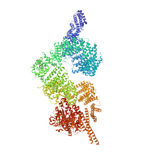
Structure of the human ATM kinase and mechanism of Nbs1 binding.

Warren, C.Pavletich, N.P.

(2022) Elife 11

  • DOI: https://doi.org/10.7554/eLife.74218
  • Primary Citation Related Structures: 
    7SIC, 7SID

  • PubMed Abstract: 

    DNA double-strand breaks (DSBs) can lead to mutations, chromosomal rearrangements, genome instability, and cancer. Central to the sensing of DSBs is the ATM (Ataxia-telangiectasia mutated) kinase, which belongs to the phosphatidylinositol 3-kinase-related protein kinase (PIKK) family. In response to DSBs, ATM is activated by the MRN (Mre11-Rad50-Nbs1) protein complex through a poorly understood process that also requires double-stranded DNA. Previous studies indicate that the FxF/Y motif of Nbs1 directly binds to ATM, and is required to retain active ATM at sites of DNA damage. Here, we report the 2.5 Å resolution cryo-EM structures of human ATM and its complex with the Nbs1 FxF/Y motif. In keeping with previous structures of ATM and its yeast homolog Tel1, the dimeric human ATM kinase adopts a symmetric, butterfly-shaped structure. The conformation of the ATM kinase domain is most similar to the inactive states of other PIKKs, suggesting that activation may involve an analogous realigning of the N and C lobes along with relieving the blockage of the substrate-binding site. We also show that the Nbs1 FxF/Y motif binds to a conserved hydrophobic cleft within the Spiral domain of ATM, suggesting an allosteric mechanism of activation. We evaluate the importance of these structural findings with mutagenesis and biochemical assays.


  • Organizational Affiliation
    • Structural Biology Program, Memorial Sloan Kettering Cancer Center, New York, United States.

Macromolecule Content 

  • Total Structure Weight: 703.32 kDa 
  • Atom Count: 44,484 
  • Modeled Residue Count: 5,546 
  • Deposited Residue Count: 6,112 
  • Unique protein chains: 1

Macromolecules

Find similar proteins by:|  3D Structure
Entity ID: 1
MoleculeChains  Sequence LengthOrganismDetailsImage
Serine-protein kinase ATM
A, B
3,056Homo sapiensMutation(s): 0 
Gene Names: ATM
EC: 2.7.11.1
UniProt & NIH Common Fund Data Resources
Find proteins for Q13315 (Homo sapiens)
Explore Q13315 
Go to UniProtKB:  Q13315
PHAROS:  Q13315
GTEx:  ENSG00000149311 
Entity Groups
Sequence Clusters30% Identity50% Identity70% Identity90% Identity95% Identity100% Identity
UniProt GroupQ13315
Sequence Annotations
Expand
Reference Sequence

Experimental Data & Validation

Experimental Data

  • Method: ELECTRON MICROSCOPY
  • Resolution: 2.51 Å
  • Aggregation State: PARTICLE 
  • Reconstruction Method: SINGLE PARTICLE 
EM Software:
TaskSoftware PackageVersion
MODEL REFINEMENTPHENIX
RECONSTRUCTIONRELION

Structure Validation

View Full Validation Report


Entry History 

& Funding Information

Deposition Data


Funding OrganizationLocationGrant Number
National Institutes of Health/National Cancer Institute (NIH/NCI)United States5F32CA247320
National Institutes of Health/Eunice Kennedy Shriver National Institute of Child Health & Human Development (NIH/NICHD)United StatesCA008748

Revision History  (Full details and data files)

  • Version 1.0: 2022-02-02
    Type: Initial release
  • Version 1.1: 2024-06-05
    Changes: Data collection