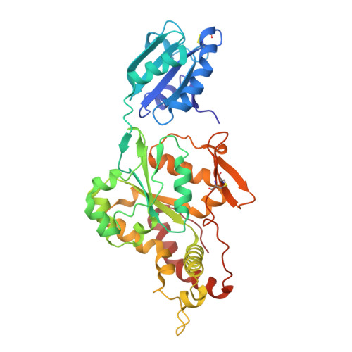
7SCD

Ternary complex of fixed-arm Trx-3ost5 (I299E) with 8mer-1 octasaccharide substrate and co-factor product PAP


Experimental Data Snapshot

  • Method: X-RAY DIFFRACTION
  • Resolution: 2.90 Å
  • R-Value Free: 0.252 
  • R-Value Work: 0.208 
  • R-Value Observed: 0.210 

Starting Model: experimental
View more details

wwPDB Validation   3D Report Full Report


Ligand Structure Quality Assessment 


This is version 1.2 of the entry. See complete history


Literature

Structural and Substrate Specificity Analysis of 3-O-Sulfotransferase Isoform 5 to Synthesize Heparan Sulfate

Wander, R.Kaminski, A.M.Wang, Z.Stancanelli, E.Xu, Y.Pagadala, V.Li, J.Krahn, J.M.Pham, T.Q.Liu, J.Pedersen, L.C.

(2021) ACS Catal 11: 14956-14966


Macromolecules
Find similar proteins by:  (by identity cutoff)  |  3D Structure
Entity ID: 1
MoleculeChains Sequence LengthOrganismDetailsImage
Thioredoxin 1,Heparan sulfate glucosamine 3-O-sulfotransferase 5372Escherichia coli K-12Homo sapiens
This entity is chimeric
Mutation(s): 1 
Gene Names: trxAfipAtsnCb3781JW5856HS3ST53OST5HS3OST5
EC: 2.8.2.23
UniProt & NIH Common Fund Data Resources
Find proteins for P0AA25 (Escherichia coli (strain K12))
Explore P0AA25 
Go to UniProtKB:  P0AA25
Find proteins for Q8IZT8 (Homo sapiens)
Explore Q8IZT8 
Go to UniProtKB:  Q8IZT8
PHAROS:  Q8IZT8
GTEx:  ENSG00000249853 
Entity Groups  
Sequence Clusters30% Identity50% Identity70% Identity90% Identity95% Identity100% Identity
UniProt GroupsP0AA25Q8IZT8
Sequence Annotations
Expand
  • Reference Sequence
Oligosaccharides

Help

Entity ID: 2
MoleculeChains Length2D Diagram Glycosylation3D Interactions
2-deoxy-6-O-sulfo-2-(sulfoamino)-alpha-D-glucopyranose-(1-4)-beta-D-glucopyranuronic acid-(1-4)-2-deoxy-6-O-sulfo-2-(sulfoamino)-alpha-D-glucopyranose-(1-4)-beta-D-glucopyranuronic acid-(1-4)-2-deoxy-6-O-sulfo-2-(sulfoamino)-alpha-D-glucopyranose-(1-4)-beta-D-glucopyranuronic acidB [auth C]6N/A
Small Molecules
Ligands 1 Unique
IDChains Name / Formula / InChI Key2D Diagram3D Interactions
A3P (Subject of Investigation/LOI)
Query on A3P

Download Ideal Coordinates CCD File 
C [auth A]ADENOSINE-3'-5'-DIPHOSPHATE
C10 H15 N5 O10 P2
WHTCPDAXWFLDIH-KQYNXXCUSA-N
Experimental Data & Validation

Experimental Data

  • Method: X-RAY DIFFRACTION
  • Resolution: 2.90 Å
  • R-Value Free: 0.252 
  • R-Value Work: 0.208 
  • R-Value Observed: 0.210 
  • Space Group: P 21 21 21
Unit Cell:
Length ( Å )Angle ( ˚ )
a = 55.84α = 90
b = 58.82β = 90
c = 153.11γ = 90
Software Package:
Software NamePurpose
PHENIXrefinement
HKL-2000data reduction
HKL-2000data scaling
PHASERphasing

Structure Validation

View Full Validation Report


Ligand Structure Quality Assessment 


Entry History & Funding Information

Deposition Data


Funding OrganizationLocationGrant Number
National Institutes of Health/National Institute of Environmental Health Sciences (NIH/NIEHS)United States1ZIA-ES102645
National Institutes of Health/National Heart, Lung, and Blood Institute (NIH/NHLBI)United StatesHL094463
National Institutes of Health/National Heart, Lung, and Blood Institute (NIH/NHLBI)United StatesHL144970
National Institutes of Health/National Institute of General Medical Sciences (NIH/NIGMS)United StatesGM128484
National Institutes of Health/National Institute of General Medical Sciences (NIH/NIGMS)United StatesGM134738
National Institutes of Health/National Heart, Lung, and Blood Institute (NIH/NHLBI)United StatesHL139197

Revision History  (Full details and data files)

  • Version 1.0: 2022-01-19
    Type: Initial release
  • Version 1.1: 2023-10-18
    Changes: Data collection, Refinement description
  • Version 1.2: 2024-10-30
    Changes: Structure summary