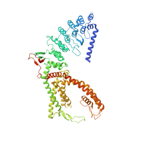
7S88

Open apo-state cryo-EM structure of human TRPV6 in glyco-diosgenin detergent


Experimental Data Snapshot

  • Method: ELECTRON MICROSCOPY
  • Resolution: 2.69 Å
  • Aggregation State: PARTICLE 
  • Reconstruction Method: SINGLE PARTICLE 

Starting Model: experimental
View more details

wwPDB Validation   3D Report Full Report


This is version 1.1 of the entry. See complete history


Literature

Structural mechanisms of TRPV6 inhibition by ruthenium red and econazole.

Neuberger, A.Nadezhdin, K.D.Sobolevsky, A.I.

(2021) Nat Commun 12: 6284-6284

  • DOI: https://doi.org/10.1038/s41467-021-26608-x
  • Primary Citation of Related Structures:  
    7S88, 7S89, 7S8B, 7S8C

  • PubMed Abstract: 

    TRPV6 is a calcium-selective ion channel implicated in epithelial Ca 2+ uptake. TRPV6 inhibitors are needed for the treatment of a broad range of diseases associated with disturbed calcium homeostasis, including cancers. Here we combine cryo-EM, calcium imaging, and mutagenesis to explore molecular bases of human TRPV6 inhibition by the antifungal drug econazole and the universal ion channel blocker ruthenium red (RR). Econazole binds to an allosteric site at the channel's periphery, where it replaces a lipid. In contrast, RR inhibits TRPV6 by binding in the middle of the ion channel's selectivity filter and plugging its pore like a bottle cork. Despite different binding site locations, both inhibitors induce similar conformational changes in the channel resulting in closure of the gate formed by S6 helices bundle crossing. The uncovered molecular mechanisms of TRPV6 inhibition can guide the design of a new generation of clinically useful inhibitors.


  • Organizational Affiliation

    Department of Biochemistry and Molecular Biophysics, Columbia University, New York, NY, USA.


Macromolecules
Find similar proteins by:  (by identity cutoff)  |  3D Structure
Entity ID: 1
MoleculeChains Sequence LengthOrganismDetailsImage
Transient receptor potential cation channel subfamily V member 6
A, B, C, D
683Homo sapiensMutation(s): 0 
Gene Names: TRPV6ECAC2
Membrane Entity: Yes 
UniProt & NIH Common Fund Data Resources
Find proteins for Q9H1D0 (Homo sapiens)
Explore Q9H1D0 
Go to UniProtKB:  Q9H1D0
PHAROS:  Q9H1D0
GTEx:  ENSG00000165125 
Entity Groups  
Sequence Clusters30% Identity50% Identity70% Identity90% Identity95% Identity100% Identity
UniProt GroupQ9H1D0
Sequence Annotations
Expand
  • Reference Sequence
Small Molecules
Ligands 4 Unique
IDChains Name / Formula / InChI Key2D Diagram3D Interactions
PCW
Query on PCW

Download Ideal Coordinates CCD File 
AB [auth C]
DB [auth D]
EB [auth D]
FA [auth B]
G [auth A]
AB [auth C],
DB [auth D],
EB [auth D],
FA [auth B],
G [auth A],
GA [auth B],
JA [auth B],
JB [auth D],
KA [auth B],
M [auth A],
N [auth A],
PA [auth C],
PB [auth D],
Q [auth A],
QB [auth D],
R [auth A],
VA [auth C],
WA [auth C],
Z [auth B],
ZA [auth C]
1,2-DIOLEOYL-SN-GLYCERO-3-PHOSPHOCHOLINE
C44 H85 N O8 P
SNKAWJBJQDLSFF-NVKMUCNASA-O
POV
Query on POV

Download Ideal Coordinates CCD File 
AA [auth B]
BA [auth B]
BB [auth D]
CA [auth B]
CB [auth D]
AA [auth B],
BA [auth B],
BB [auth D],
CA [auth B],
CB [auth D],
DA [auth B],
EA [auth B],
GB [auth D],
H [auth A],
HA [auth B],
I [auth A],
IA [auth B],
J [auth A],
K [auth A],
KB [auth D],
L [auth A],
LB [auth D],
MA [auth C],
MB [auth D],
NB [auth D],
O [auth A],
OB [auth D],
P [auth A],
QA [auth C],
RA [auth C],
SA [auth C],
T [auth A],
TA [auth C],
UA [auth C],
W [auth B],
XA [auth C],
YA [auth C]
(2S)-3-(hexadecanoyloxy)-2-[(9Z)-octadec-9-enoyloxy]propyl 2-(trimethylammonio)ethyl phosphate
C42 H82 N O8 P
WTJKGGKOPKCXLL-PFDVCBLKSA-N
Y01
Query on Y01

Download Ideal Coordinates CCD File 
E [auth A]
F [auth A]
FB [auth D]
HB [auth D]
IB [auth D]
E [auth A],
F [auth A],
FB [auth D],
HB [auth D],
IB [auth D],
LA [auth C],
NA [auth C],
OA [auth C],
S [auth A],
V [auth B],
X [auth B],
Y [auth B]
CHOLESTEROL HEMISUCCINATE
C31 H50 O4
WLNARFZDISHUGS-MIXBDBMTSA-N
CA
Query on CA

Download Ideal Coordinates CCD File 
U [auth A]CALCIUM ION
Ca
BHPQYMZQTOCNFJ-UHFFFAOYSA-N
Experimental Data & Validation

Experimental Data

  • Method: ELECTRON MICROSCOPY
  • Resolution: 2.69 Å
  • Aggregation State: PARTICLE 
  • Reconstruction Method: SINGLE PARTICLE 
EM Software:
TaskSoftware PackageVersion
MODEL REFINEMENTPHENIX
RECONSTRUCTIONcryoSPARC3.1

Structure Validation

View Full Validation Report


Entry History & Funding Information

Deposition Data


Funding OrganizationLocationGrant Number
National Institutes of Health/National Cancer Institute (NIH/NCI)GermanyR01 CA206573
National Institutes of Health/National Institute of Neurological Disorders and Stroke (NIH/NINDS)GermanyR01 NS083660
National Institutes of Health/National Institute of Neurological Disorders and Stroke (NIH/NINDS)GermanyR01 NS107253
National Science Foundation (NSF, United States)Germany1818086
German Research Foundation (DFG)Germany464295817

Revision History  (Full details and data files)

  • Version 1.0: 2021-11-17
    Type: Initial release
  • Version 1.1: 2024-06-05
    Changes: Data collection, Refinement description